Skip navigation

The NaturePlus Forums will be offline from mid August 2018. The content has been saved and it will always be possible to see and refer to archived posts, but not to post new items. This decision has been made in light of technical problems with the forum, which cannot be fixed or upgraded.

We'd like to take this opportunity to thank everyone who has contributed to the very great success of the forums and to the community spirit there. We plan to create new community features and services in the future so please watch this space for developments in this area. In the meantime if you have any questions then please email:

Fossil enquiries: esid@nhm.ac.uk
Life Sciences & Mineralogy enquiries: bug@nhm.ac.uk
Commercial enquiries: ias1@nhm.ac.uk

1 2 3 4 Previous Next

Curator of Micropalaeontology's blog

52 Posts tagged with the palaeontology tag
0

On the 18th and 19th of November we welcomed over 150 micropalaeontologists who travelled from as far away as New Zealand for a conference at the Museum. Over 50 of them were given tours behind the scenes and 45 micropalaeontology posters were displayed. Thank you to fellow conference hosts and Museum scientists Tom Hill and Steve Stukins who have written this guest blog to describe the significance of these two busy days/weeks/months for micropalaeontology at the Museum:


IMG_2733_blog.jpg

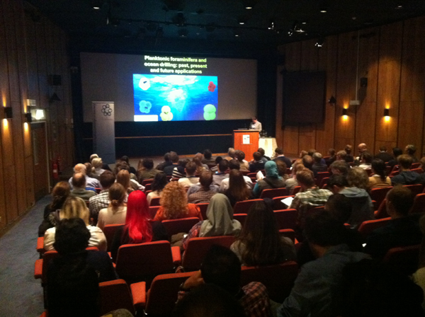
The stage set for the start of the micropalaeontology conference at the Museum


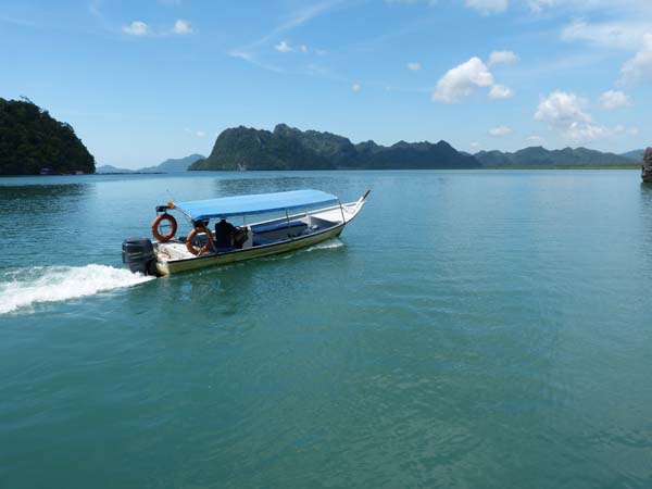
On Monday 18th and Tuesday 19th November 2013, the Micropalaeontology Unit hosted a conference at the Museum on behalf of The Micropalaeontological Society under the theme Micropalaeontology and the IODP: Past, Present and Future Applications. IODP stands for the International Ocean Drilling Programme and is an international marine research collaboration that explores Earth's history using ocean-going research platforms, for example the JOIDES Resolution research vessel. These platforms drill cores through the seafloor to recover the layers of sediment and rock that have accumulated at the bottom of the oceans.


The conference was a resounding success, with 150 delegates registering and contributing. We even had to close registration a couple of weeks beforehand as we reached capacity. The event included a number of keynote speakers who specialise in research on IOPD samples, numerous presentations and posters covering all aspects of micropalaeontology and tours behind the scenes.

IMG_0717_blog.jpg

Conference delegates enjoying the poster session


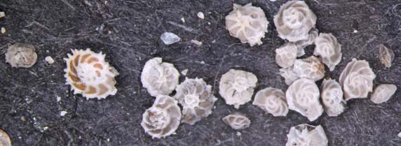
Micropalaeontology is an integral part of IODP studies and as a result micropalaeontologists will be found on all IODP cruises. As the amount of sediment or rock extracted by coring is often relatively small, microfossils provide an ideal route through which scientists can:

  • study the age of the sediment under investigation
  • reveal the climatic conditions that prevailed at the time of sediment deposition

This is because hundreds, thousands, indeed millions of microfossils may be preserved in a layer of sediment only a few millimetres thick. As a result, micropalaeontology is at the forefront of IODP and climate change research.


IMG_2723_blog.jpg

Keynote speaker Prof Bridget Wade from University College London


The international relevance and importance of micropalaeontology in ocean research was underlined by the fact that we hosted delegates from across the world including America, Canada, France, Germany, the Netherlands, New Zealand, Switzerland and Tunisia, to name a few. The diversity in delegates was complemented by the similar diversity in micropalaeontology projects that were discussed through both oral and poster presentations.


One of the research topics often discussed was how micropalaeontologists can extract detailed geochemical signals from microfossils to quantify past oceanic and climatic conditions. If such high resolution analysis is applied to an ocean core, it is then possible to reconstruct how oceanic conditions (which in turn reflect climate) have changed over millions of years. Such studies are essential if scientists are to understand how our Earth responds to climate change (whether it be due to natural or human-induced change).


IMG_0774_blog.jpg
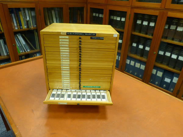
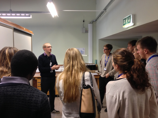
Steve Stukins demonstrating the John Williams Index of Palaeopalynology to a group of M.Sc. students studying Applied and Petroleum Micropalaeontology at the University of Birmingham


The conference also provided a unique opportunity to publicise the quality and quantity of the micropalaeontology collections housed at the Museum to a large specialist audience. Steve and Giles provided tours through the collections to over 50 micropalaeontologists including the new intake of MSc students studying micropalaeontology at Birmingham University.


Numerous new projects to work on our collections and potential new collaborations have resulted from these tours and from discussions we have had during the conference. From the feedback that we received, it seems that members of the Micropalaeontological Society also found the conference a great success. If you are interested to learn more details, the abstract volume for the TMS2013 talks is now available on-line.


The conference would not have been such a great success had it not been for the generosity of our sponsors. We would therefore like to thank The Micropalaeontological Society, The UK-IODP, The Geological Society, AASP – The Palynological Society, Neftex, Olympus, Petrostrat and Beta Analytic for their kind support during this event.

0

My diary this week is illustrated by an item from behind the scenes at the Museum for each day. The week's events include:

  • an offer of an important historical collection for sale
  • work towards digitising an amazing collection of marine plankton pictures
  • hosting an old friend from Brazil
  • a recent donation appearing in a publication
  • preparation for a session on the scanning electron microscope


Monday

Below is an example from our collection of a slide made by the famous slide mounter Charles Elcock in 1880. Slides made by Elcock fetch as much as £350 on the open market so it was very exciting to be approached by a dealer to see if we wanted to acquire his foraminiferal collection and archive. Unfortunately the asking price was significantly more than we can afford.

Elcock_DSC3569.jpgA slide from our collections made by Charles Elcock in 1880.


We regularly buy specimens, and in fact some of my colleagues recently purchased specimens at the Munich Rock and Fossil Show. As far as I know, the last item bought for the micropalaeontology collection was a conodont animal back in the late 1980s.


At the very least this collection offer makes me realise the monetary value placed on historical items in the collection that I look after.


Tuesday

Below is a scanning electron microscope image of a coccolith, part of a collection of over 6,000 images taken by my former colleague Dr Jeremy Young and his collaborators.

207-23-syra. molischii_SU_blog.jpgScanning electron microscope image of the coccolith Syracosphaera molischii.


Recently graduated micropalaeontology Masters student Kelly Smith is visiting today to help us work up the data on this collection so that we can make information and images available via our website. Because their distribution is controlled largely by temperature, coccoliths in ancient sediments have been used to provide details of past climate change.


Coccoliths make their tiny calcareous shells by precipitating calcium carbonate from seawater so their present-day distribution may have been affected by acidification of the oceans relating to the burning of fossil fuels.


Wednesday

In 20 years at the Museum I have met a lot of people from all over the world, some of whom have visited regularly and subsequently become good friends. I first met Prof. Dermeval do Carmo of the University of Brasilia, Brazil, shortly after I arrived at the Museum. We have enjoyed a long collaboration that has included working together on publications, teaching collections management in Brazil, fieldwork and even holidays. He is visiting with a large, empty suitcase to to pick up the last of the scientific papers identified as duplicate by some of our volunteers.


11563_large.jpg

The Brazilian Cretaceous non-marine ostracod Pattersoncypris micropapillosa.


Today's picture is the exceptionally preserved Brazilian Cretaceous non-marine ostracod Pattersoncypris micropapillosa. Dermeval has recently reclassified the genus under the name Harbinia and is on his way to China to look at the type material for this genus. Species related to Pattersoncypris/Harbinia are used to provide information on rock ages and environments of deposition for oil exploration offshore Brazil and W Africa. For more details about their evolutionary significance, see my post on What microfossils tell us about sex in the Cretaceous.


Thursday

Today's scanning electron microscope image is the holotype of a new species of planktonic foraminifera Dentoglobigerina juxtabinaiensis, donated last summer and published last month in the Journal of Foraminiferal Research by University of Leeds PhD student Lyndsey Fox and her supervisor Prof. Bridget Wade.


The Museum collection contains many type specimens that ultimately define the concept for each species. As a result, types are some of our most requested specimens by visitors or for loan.


Fig 7.1c (holotype) u1337a 42xcc_blog.jpg

The holotype of a new species of planktonic foraminifera, Dentoglobigerina juxtabinaiensis.


This species is Miocene in age (roughly 13.5-17 million years old) and was recovered from an International Ocean Discovery Programme core taken from the Pacific Ocean near the equator. Foraminifera, like the coccoliths mentioned earlier, are important indicators of ocean condition and climate, so this unusually well-preserved material is an important contribution to their study. Now the paper has been published I am able to register the details in our database and these will go live on our website in a couple of weeks.


Friday

I have a scanning electron microscope session booked next week as I have been approached by a colleague at the British Geological Survey who is preparing a chapter for a field guide on Jurassic ostracods. He would like some images of some of our ostracod type specimens from the Kimmeridge Clay Formation of Dorset.

HULL631_Mandelstamia_maculata_cgmq_107_blog.jpg

The ostracod Mandelstamia maculata from the Jurassic Kimmeridge Clay of Dorset.


Each specimen must be carefully removed from the slides that house them using a fine paint brush and glued to a small aluminium stub about 1cm in diameter. These will be coated in a fine layer of gold-palladium before being placed in the scanning electron microscope chamber next week for photography.


It proves very difficult to identify the relevant specimens as the original publication is pre-scanning electron microscope times and the images taken down a binocular microscope are less than clear. Publishing new, clearer illustrations of each of these type specimens will add considerable scientific value to our collections.


This selection of specimens has hopefully shown the historical, scientific and monetary value of our collections while showing that they are also relevant to important topical issues such as climate change and oil exploration.

1

A few weeks ago the Center for the Future of Museums blogged on how to get your museum blog widely read, sadly stating that writing for a niche audience like micropalaeontology is unlikely to be successful. As a reply I thought I would share my own experiences on what has worked for me and what hasn't, with nine tips for museum bloggers.


Choose an eye-catching title and subject

The title and subject ultimately convince the reader whether to visit your post or not. I agree with the Center for the Future of Museums that blogs with titles and subjects that are of general interest will be more widely read, as the post asking Do we need specialist curators? has shown. However, it is also possible to write about your specialist collection and make it relevant to a general audience, such as What microfossils tell us about early humans in Britain or When microfossils meet dinosaurs. Other more specific posts describing collections, databasing or risk management have not been so well read.


Utilise social media to advertise

I quickly realised that there was no point in posting and just hoping that people will automatically find what you have written. Posts tweeted by @NHM_London, which currently has over 400,000 followers, receive significantly more hits than others. We started the @NHM_Micropalaeo Twitter feed to provide micropalaeontology news from the Museum and to advertise posts from this blog. Other relevant advertisement vehicles have been sites such as Facebook, Reddit or LinkedIn, while #AskACurator day on twitter was also a great opportunity to publicise our collections by highlighting previously published blog posts.


Build links with other bloggers and webmasters

Link regularly to associated websites and write to the site owners to let them know that you have done so. I link regularly to the Geological Curators' Group, Forams.eu and The Micropalaeontological Society and they have all provided reciprocal links. Other sites such the Museum website, Focus Magazine and blogger Tony Edger have provided links to my writing, prolonging the reading life for some posts way beyond the point when they are not visible on the blog front page. 


Link to other sites clearly

The visibility of your blog to search engines such as Google is significantly enhanced if you link via a string of text that describes the link. For example, it is best to link to The Micropalaeontological Society rather than writing 'click here'.


Run your own email distribution list

Most sites, like this one, have the option to subscribe and receive updates when new posts go live. This works reasonably well if a lot of people make the effort to create an account and log on to follow your posts. However, most readers I have spoken to do not do this. I have lost count how many times I have heard people say 'I like your blog but I haven't looked at it recently'. I have set up a mailing list based on people I regularly deal with and send a message out every time I post a new blog. Posting blog links to relevant academic listservers has also been successful in generating additional readers.


Write for an audience

I write all posts as if I am explaining to my mother or mother-in-law, but at the same time making the post interesting to experts in the field of micropalaeontology wanting the latest news from the Museum. I feel confident that I am reaching my target audiences as I have been pleased to receive feedback  from a wide range of readers, including:

  • university academics
  • students
  • school teachers
  • amateur micropalaeontologists
  • members of the public


Think carefully about your reasons for blogging

In my first post I gave the following reason for starting this blog: 

In this age of austerity, I believe that we should be highlighting the good news coming from the Museum so that the applications and relevance of our collections, including those from micropaleontology, are brought to people’s attention.

Even when writing more general posts like this one, I have this theme in my head while writing so it is always possible to include information about our collections and their relevance.


Get your timing right

There is no point in publishing a Microfossil Christmas card blog piece on Christmas Eve and hoping that lots of people will be logging on to read it. Similarly if you are going to tweet about your latest post it is best to do it at at time when most people are likely to read it. My successful post on How to become a curator? was timed to coincide with half term and a gallery exercise called 'curious curators'. Another post went live to coincide with the opening of our Treasures Gallery at the Museum.


Include a good balance of personal narrative

How much you write about yourself and your feelings depends on your writing style and subject of your blog. My blog highlights the collections and their use and not myself, so I sometimes feel that I do not include enough of my own personal story. Posts where I have shown how passionate I am about collections and collecting like my post on 'How to become a curator have been well received though.


Post regularly

If you don't get overnight success then don't give up. It takes time to gain a following, build relationships with other bloggers, webmasters or fellow Tweeters. By definition the more often you post, the more hits you'll get. People are more likely to follow or keep checking a blog that is active.


It has been hard work but as a curator I feel that blogging about my collections has had a major impact on their profile and I would encourage any curator to do the same. In summary, if you write interesting material that gets tweeted and retweeted around the internet then people will read it, whether you are writing about 'niche collections' or not.

0

This is my 50th blog post, so I thought I would look back and make a list of benefits that have come directly from blogging about my job and the collections in my care. These include an enhanced profile of the collection, help with collections management, fundraising, research collaboration offers and an enhanced personal profile.


There are probably more that can't be directly measured but here are 20 to be going on with:


Press coverage

1. The post on microfossil Christmas cards inspired an article in the Independent in December 2012.

2. The item on specialist curators was published in full on the Museums Association (MA) website.

3. The same post was one of the most read for 2012 on the MA website.

4. The Guardian used my post on specialist curators as a basis for an on-line poll.

5. The first paragraph of my post on volunteers was quoted in the Museums Journal under the title 'Best of Blogs'.

6. Images of slides from the collection were reproduced on the ScienceFocus website.


Collection management

7. I have been able to answer a number of internal and external enquiries by providing a link to blog posts.

8. A researcher from University College London has offered some grant money towards CT-scanning some of our holotype specimens.

9. Some readers have provided information to enhance the collections by identifying unnamed specimens.

10. I have been able to expand my knowledge about some important parts of the collection that previously I knew little about.


Collection usage

11. We have had a marked increase in the number of artists using the collection.

12. Some collection images featured on the blog have been sold via the Museum's Picture Library.

13. We have had three exhibition loan requests to display microfossil-related items, including a CT scan.


My research

14. I was asked to co-author a paper following my post on virtual loans.

15. I have had a request to participate in an exciting research project on ocean acidification that includes funding for more CT scanning.

16. A high profile journal has asked me to review a microfossil-related book.


Advisory role

17. A number of people have requested career advice, with one recently accepting a job in collection management.

18. We were approached by PalaeoCast to make a podcast about micropalaeontology.

19. I have had requests for advice on starting a blog.


And finally, relating to my personal development ....


20. I feel that blogging has helped me to write faster and more concisely.


I hope you will agree that this blog has enhanced the profile of the micropalaeontology collections both within and outside the Museum. There are still plenty of interesting issues and collections to write about. Please keep reading to find out how our microfossil specimens play a major role in climate studies and how a microfossil sculpture park in China relates to our collections.

0

This week I celebrate 20 years at the Museum, and my diary has included preparing for a researchers' night highlighting museum science, Tweeting as part of #AskaCurator day and visiting a miniature steam railway.


Monday


Most of today has been spent preparing for Science Uncovered, our EU-funded researchers' night on Friday 27 September. The doors of the Museum will remain open after usual closing time and scientists like myself will be available to talk about our science, show specimens and chat. Presentations in the Nature Live Studio will also be held and it will be possible to book tours to areas of the Museum not normally open to the public.


P1020914_blog.jpg

This core from the Atlantic SW of Ireland represents the last major glacial period showing glacial dropstones from colder periods (left) and white sections composed almost entirely of warm water microfossils (right). The green packets and plastic sleeve maintain an oxygen free environment.


We are showing some deep sea cores taken from the Atlantic Shelf off SW Ireland through sediment that was deposited during the last glaciation. It's a great opportunity to show the key role micropalaeontology plays in quantifying and dating past climatic episodes. The core relates to periods when icebergs broke off glaciers and traversed the North Atlantic.


Tuesday


A major part of my job is dealing with enquiries about our microfossil collections and subsequently hosting visits or preparing loans. Two main collections are our most requested, the Challenger Foraminifera and the Blaschka glass models of radiolarians. Since three specimens from our Blaschka collections have been on display in our Treasures Gallery, we have had an increased number of enquiries and visitors to view the other 180 specimens that are not currently on display.


0035_blog.jpg

Image of one of our Blaschka glass radiolarian models that was widely retweeted during #AskACurator day.


Today we are showing the undisplayed Blaschka collection to an artist, last week it was a glassworker from Imperial College and later this week it is a photographer hoping to create a book of images from Blaschka collections across Europe.


Wednesday


I have spent virtually the whole day on Twitter monitoring questions and providing answers as part of #AskACurator day. Fellow curators from over 500 different museums and 35 different countries have been fielding questions over Twitter causing the hashtag to trend. At one stage it was globally the second most discussed subject on Twitter.


I answered questions like:


'Do you require a masters degree to become a curator?'

'Which museum, other than your own, inspired you recently?' (the Foraminiferal Sculpture Park in China)

'Which specimens in your collection give you goosebumps when you see them?' (Blaschka glass models

'What sparked your interest to become a curator?'

'Do you need to be an obsessive to be a curator?'

'Which specimen not currently on display would you like to see being displayed?' (100 year old microfossil Christmas card).


Many of the questions I was able to expand on using links to blog posts, particularly the one entitled 'How to become a curator'. I started to reply to the 'what is a curator?' question but could not cram 'someone who cares for a collection by enhancing its documentation and storage, maintains access to it by facilitating loans, visits and exhibits and promotes its relevance by engaging with potential users' into 140 characters.The day certainly showed what a varied job we all have, how passionate we are and that one day is never the same as another.


Thursday


My colleague Steve has worked out that we had 65 new interactions (messages, favourites, retweets, new followers) during #AskACurator day yesterday as well as some more hits to this blog. However, I am saddened as I read a well known museums blog that says that the best way to reach a wide audience is to avoid niche subjects like micropalaeontology and links direct to my blog as an example. It starts me wondering if accumulating vast numbers of hits really show that a blog is successful?


A string of meetings are scheduled too; we are applying for funding for a major 3 year conservation project, the photographer arrives to discuss his project and we are finishing an application to hire a new PhD project studying traits of evolution. Microfossils are extremely useful as their fossil record is relatively complete compared to other fossil groups and collections can be made relatively easily across large geographical areas.


Friday


Earlier in the week I got to work to find two of my train mad, three year old son Pelham's Thomas the Tank Engine stickers on my socks. On Monday he is starting nursery school so today we are taking him and his younger sister Blossom to one of our favourite places, the Romney, Hythe and Dymchurch Railway in Kent. I feel that this family day is a suitable way to celebrate the 20th anniversary of my arrival at the Museum as a volunteer.


P1020924_blog.jpg

0

On Friday 27 September the doors of the Museum will remain open after usual closing time and scientists like me will be available to talk about our science, show specimens and chat at Science Uncovered, our EU funded Researchers' Night. Presentations in the Nature Live Studio will also be held and it will be possible to book tours to areas of the Museum not normally open to the public.


This year Tom, Steve and I are on the Climate Change table in Waterhouse Way demonstrating some deep sea cores taken from the Atlantic Shelf SW of Ireland. The cores were drilled through sediments representing the last ice age. Information on the distribution and composition of microfossils, allied with other scientific data, shows six 'Heinrich Events' through the last glaciation. These events are thought to relate to climate related cyclic episodes when icebergs broke off glaciers and traversed the North Atlantic.


Science Uncovered 2012 welcomed an incredible 8,523 visitors over the night who spoke to over 350 scientists. If it proves to be as successful as last year where we presented our microfossil zoo or 2011 when I was able to use a giant plasma screen to show some of my research then it promises to be an amazing night. Do come and join us if you can.


P1020918_blog.jpg

This core from the Atlantic SW of Ireland represents the last major glacial period showing glacial dropstones from colder periods (left) and white sections composed almost entirely of warm water microfossils (right). The green packets (far right) and plastic sleeve maintain an oxygen free environment and prevent mold growth on the core.

2

The Museum has been running an inspiring schools activity under the "How Science Works" strand of the National Curriculum, in which children carry out their own microfossil research project. In just over an hour, they investigate the microfossils in some Gault Clay collected from Folkestone, and learn the processes behind research including how scientists collect data, use it to solve problems and publish their findings.


Feedback from the school groups shows that it is a fun and engaging way to learn about science away from the normal classroom environment. After completing the exercise, many children indicated that they were more enthusiastic towards a career in science and some even felt that they were already scientists!

How_Science_works_sally_871X9998.jpgSally Collins and Jamie Robinson about to start a "How Science Works" session with a school group at the Museum.


The activity was developed by Sally Collins, currently part of the Museum Content Development Team, and Dr Craig Koch, while he was studying for his PhD in the Palaeontology Department. It can now be run entirely by Museum educators, while the use of commonly available kit such as washing up bowls and plastic screw topped bottles means that there is potential for holding this schools exercise in locations away from the Museum.


The 'How Science Works' activity


Before the exercise, Gault Clay from Folkestone is crushed into 1-2cm lumps while still wet, and subsequently left to dry. At the start of the class, approximately 100g is then placed in a plastic screw-topped container and boiling water added. After the container has cooled down a bit and some cold water added to bring it down to a safe temperature, it is shaken rather like a cocktail shaker.


While the sediment is soaking, there is time for the class leader to explain the exercise and how micropalaeontology is used in earth science research. A video is shown, featuring Craig and his supervisor Dr Jeremy Young on a research cruise and working in the laboratories at the Museum.


How_Science_Works_sieves_blog.jpg

Sieving sediment in the classroom.


After soaking for 15 minutes, the sediment is sieved, dried in a microwave and rehydrated before a final sieving and drying. The microwave heating and subsequent drying ensures that the process can be carried out in a short period of time.


The small microfossil residues produced are then examined under a microscope by the children and a chart is provided to help them identify the microfossils present. By analysing the geological ranges of all the species present, it is quickly possible for the children to determine the age of the clay.


This is similar to the work that a micropalaeontologist might carry out on a well site, where age information could be used by the drillers to decide to continue drilling or stop. Drilling a well too deep can be a costly mistake so sometimes the micropalaeontologist on a well site is put in a major decision-making position!


How_Science_Works_range_chart_blog.jpg

The chart used by the students to identify their microfossils and  decide on the age of the clay.


There is still time for a final class discussion of the results obtained by each group and how scientists communicate their findings by publishing their data. The session I attended recently ended with a very entertaining discussion amongst the children about how the 'peer review process' effectively makes sure that scientists' results are checked before they are published.


Teacher and student feedback


Some of the school class teachers have provided feedback, including:

'Perfectly engaging, challenging and inspiring

'The students were pretty curious and asked a lot of questions which in my opinion shows that the workshop had a stimulating effect on them.'

Children taking the exercise have also had the chance to feedback. Here is a selection of answers given when participants were asked if taking the class had affected their plans for taking science:

'I was thinking about studying science for A-level beforehand -  my visit today has reinforced that'

'Yes makes me want to study science more'

Yes, I’d love to do something related to micropalaeontology'

Yes because now I am thinking of becoming a scientist'

Yes, I will go home and purchase a microscope!'

'I will take my science lessons more seriously'

'No it has improved my desire to study science more. I am inspired.'

Epistomina_spinulifera-A17_10-slide-2_5x_crop_blog.jpg

The foraminifera Epistomena spinulifera; one of the age indicative species of microfossils in the clay.


When the children were asked to say what they 'didn’t know or never realised' some answers were:

I never realised that ...

‘you could have so much fun with clay’

'there were microfossils in clay'

'I do actually like biology'

'that rocks could be dated!'

'science facts had to go through a long process before getting published'


Finally some general comments that I think sum up the impact of the exercise nicely:

'We got to do our own experiment and discover things and come to a conclusion which made us feel like scientists'

[I enjoyed it very much] 'because it was really fun and I learnt more than I probably would in a normal science lesson'

'I enjoyed looking at the fossils from millions of years back and it makes me feel special to realise that I am the first one to see them'

[I enjoyed it very much] 'because we could be scientists and actually find the results on our own. Rather than a classroom where you are told what happens and told the results.'

How_Science_Works_range_chart_closeup.jpg


This positive feedback from previous "How Science Works" schools vists shows that it is a very successful format for learning. Unfortunately it is not currently being run by the Museum due to lack of bookings and staff resource issues. Sally Collins reports that 'we could only run it for half a class at a time due to the space limitations of the Earth Science Teaching Room and the need for everyone to be able to access sinks and electricity. The trend in the past couple of years has been for secondary schools to bring larger and larger groups.'


The exercise has been modified to use washing up bowls instead of sinks and the use of microscopes in other spaces in the Museum such as the Earth Lab mean that larger groups could be accomodated in the future.


The Gault Clay exercise is currently being run successfully at Peterborough Museum. My colleague Dr Steve Stukins has been working with Peterborough and the Natural History Museum's Real World Science Officer Hannah Pritchard to develop a similar hands-on activity using local Oxford Clay. I look forward to hearing about the Oxford Clay exercise and hope to see the successful Gault Clay schools activity running again at the Museum soon.

0

My work diary of last week, in which members of the public put a valuable part of the collection at risk with their smart phones, tiny floating snails cause a flurry of visitors and microfossils are mentioned on the Test Match Special cricket commentary(!) in a varied week for the curator of micropalaeontology.


Monday


First up is a trip across the Museum to the Nature Live Studio with some delicate specimens that will be the subject of my two public talks later in the day. We can't move large items across the Museum during opening hours and, with the galleries filled with summer visitors, this is more than sensible at present.

Nature_Live2013_3_blog.jpg

In the Nature Live Studio with host Tom Simpson - a CT-scan of a Blaschka glass radiolarian model on the screen.


All of the specimens in my care bar the one in the Treasures Gallery are housed behind the scenes, so regular visitors might not even know that we have microfossil collections. A previous head of palaeontology collections calculated that we have as few as 0.001 per cent of our fossil collections on display.


If you haven't been to one yet, the Nature Live events are a great way to bring these parts of our collections out for the public and allow us to talk about our science. The incredibly delicate and unique Blaschka Glass models of radiolarian microfossils are always a big hit, but we have to ask a smart phone-brandishing throng of children and their parents to move away from the specimens after the first show as a mother leans over the barrier and takes a picture on her phone from right above the specimen. We add two extra barriers for the second show!


Tuesday


I've had an enquiry from The Geological Magazine asking me to review a book that I have almost finished reading. I have to think carefully about saying yes or no. Receiving a free copy is the usual bonus for undertaking these tasks but, as I have a copy already, dedicating a lot of time to a review does not seem so appealing.


I decide that I shall send the extra copy to my student in Malaysia but I think I will wait until after she has finished writing up her MSc thesis. Her first chapter arrives today for comment as do some proofs for a book chapter on microfossil models that I have written. Much of the day is spent checking these and providing additional information requested by the editors of the book. It will be about the history of study of microfossils and will have an image of one of our microscope slides on the cover.


Wednesday


The galleries are packed with summer visitors but it is relatively quiet behind the scenes with many staff on annual leave, away on study trips, conferences or fieldwork.


This quieter period is a good time to catch up on some of the documentation backlog so today I finish documenting a new donation, continue to work on a large dataset relating to specimens from the Challenger Collection, and register 30 Former BP Microfossil Collection specimens that are due to go out on loan to the USA.


I spent my first 6 years at the Museum on a temporary contract curating the Former BP Microfossil Collection so it is always satisfying to see it being used by scientists. We have big plans for this collection in the future. However, I feel that I will need to do more than wait for a rare quiet day if I am to meet my part of the databasing targets set by the Museum. We plan to have details of 20 million of our specimens on our website within 5 years.


Thursday


An enquiry has come in this week about our heteropod collection. These are tiny planktonic gastropods, or literally floating snails. They are of great interest to scientists looking to quantify the effects of ocean acidification because they secrete their shells of calcium carbonate directly from the seawater that they lived in.


Measurement of carbon and oxygen isotopes from fossil examples can give details of the composition of ancient oceans and help to quantify changes over time. I mention the enquiry to staff in the Life Sciences Department and three visitors arrive to look at our collection within a day, including two from the British Antarctic Survey looking to develop projects on ocean acidification.


Friday


It is back to documentation again, a task I often save for days when the cricket is on. I am amazed when I hear dinoflagellates mentioned during the Test Match Special commentary. Dinoflagellates are protists that are a major consituent of modern and fossil plankton. We have thousands of glass slides of them here at the Museum within the micropalaeontology collections.


straussii_cookii_montage.jpgNew species of Australian Jurassic dinoflagellate Meiogonyaulax straussii (1-4) and Valvaeodinium cookii (16-20) published by Mantle and Riding (2012). Images courtesy of Dr Jim Riding, British Geological Survey.


A regular visitor to our collections who works at the British Geological Survey in Nottingham has described two new species of Jurassic microfossil from the NW Shelf of Australia and named them strausii and cookii after the former and current England cricket captains and Ashes-winning opening batsmen. It causes much merriment in the Museum microfossil team as another former England Captain, Michael Vaughan, remarks on the radio that they look rather like omelettes.


Cricket is the theme for today as I attend a lunchtime retirement party for a former cricketing colleague at the Science Museum next door. I leave a colleague to take a visitor to lunch but come back to find that he has gone home sick and the visitor is still here with a list of requests that account for the rest of the day...

0

According to January 2013 figures on their websites, the American Museum of Natural History (AMNH) has more than 32 million specimens, the Smithsonian Institution in Washington over 126 million  and the Natural History Museum over 70 million. Do we really know how many specimens we have here at the Museum? Are these figures meaningful and does it matter if we haven't estimated correctly?


When I consider the collections in my care I often have a chuckle about these figures and wonder if I could ever even get close to estimating the actual number of specimens in my collection. Take the jars and bottles below for example; there are literally hundreds of thousands of microfossils in there.


In this post I take you through a recent calculation to estimate the number of items we have in our micropalaeontology collection, and conclude that understanding how these collection sizes have been estimated is essential in deciding how to manage them.


P1020725_blog.jpgBottles of microfossil residues containing literally uncountable numbers of specimens.


It is relatively easy to make a quick and accurate size calculation for some parts of the microfossil collection. Slides are housed in standard cabinets holding 105 drawers that each hold 55 slides. The 24 standard cabinets in the Heron-Allen Microfossil Library therefore contain roughly 138,600 slides if they are full. By similar calculations, the Former Aberystwyth University Microfossil Collection contains a total of about 60,000 slides and the Former BP Microfossil Collection 300,000.


However, some cabinets are not completely full so we estimated percentage of expansion space and scaled down the figures accordingly. The total number of microfossil slides in the entire collection is estimated to be about 550,000.


But this is an estimate for the number of slides, not specimens. One slide, like the residue bottle, may contain 10s, 100s or even 1000s of specimens. Is it worth counting all of these? Probably not. You'd be there forever. Obviously when calculating the 70 million specimen figure, these vast numbers of additional specimens have not been taken into consideration otherwise the microfossil collection would have accounted for a large percentage of the total 70 million figure and perhaps even surpassed it!


_DSC3563_blog.jpg

This slide was counted as a single item in our size estimate for the microfossil collection. Each square contains a different species and multiple specimens are present, so these counts could legitimately be added to our total number of specimens for the collection.


It would appear that by counting slides and not the specimens on them, we are making the microfossil collection and hence the Museum collection appear smaller than it is. So does this matter?


This really depends on how you use the information. I think it is fine to give estimated figures like we do on the Museum website as it gives members of the public an idea of the vast size of the collection. On the other hand, if you use these figures to make decisions on how to allocate resources to the collection, then it becomes really important to account for the way in which the data is generated.


It wouldn't be right for example, to decide how much funding to give a museum relative to another one based on figures like these, without knowing how they had been generated. It's probably unwise to take too much notice of website details of the relative sizes of collections at the AMNH, Smithsonian and Natural History Museum, as the data has almost certainly been gathered in a different way by each institution.

PF_70832_Various_Foraminifera_Christmas_1921.jpgAnother slide with multiple specimens that counts for a single item within the 550,000 slides in the microfossil collection. The story behind this slide can be found in my Microfossil Christmas Card post.


If we have 70 million specimens in the Museum, and just over half a million in the microfossil collection, which is looked after by one curator, it would, on average, suggest that we need 140 curators to manage the entire collection. The actual figure is closer to 100. Taking these figures literally would therefore suggest that I am doing well to only have to manage half a million specimens!


Of course it is not that simple. Data derived from other parts of the Museum collection are not comparable. A tray of 100 identical sharks teeth for example would have been counted as 100 individual specimens, whereas the squared microfossil slide shown above would have counted as an individual item. Other parts of the collection might appear to require more management resources, until they are compared on an equal basis by separating out curatorial units sometimes referred to as 'collection lots'. The tray of 100 sharks teeth in this instance would count as one collection lot.


It would be wrong to suggest that collection size estimates are the only factors taken into consideration when deciding how to allocate resources across a vast collection like ours. Monetary value, state of conservation, suitability for display, visitor and loan demand, educational, scientific and historical significance are also taken into account. 


I would say that 70 million is probably an under-estimate of the size of the Museum collection if you take into consideration the 'microfossil factor' of collections where there are simply uncountable numbers of specimens within collection lots. I don't think we will ever come to a meaningful total if we attempt to count individual specimens.


However, it is vital that we are consistent in how we interpret the figures derived from our own collection, especially if we use them to help make decisions on how to manage it in the future. An estimation of the number of lots rather than specimens would help towards this.

1

For obvious reasons I have always wanted to do fieldwork on Pulau Langgun, one of the islands that make up the Langkawi Islands off the far NW coast of the Malaysian Peninsula. In my last post I described the difficulties of interpreting the geology of the Malaysian Peninsula and how we were attempting to use conodont microfossils to answer some of the questions by dating the isolated rock exposures.


We travelled to Langkawi Island to sample conodonts from the most complete exposure of rocks in the region as it will act as a reference section for our student Atilia Bashardin and help to interpret the isolated rock exposures on the mainland. The islands are also where the conodont Panderodus langkawiensis was first described and this species was a target for our collecting.


P1010957.JPG


For two days our field vehicle (above) left us on the coastline early in the morning. The unfavourable tides meant that we only had three hours on the section before our boat had to come and pick us up.


P1010865.JPG


The early morning trip to the section was amazingly picturesque as we motored through mangroves and past imposing forested limestone crags. On the first morning we encountered dolphins as we moved out into the open water.


P1020105.JPG


This was our first sight of the study section from the sea. Initially it was difficult to see any rock exposures as they are hidden behind the margins of the forest that encroaches the beach. We landed at the end of the pier that you can see to the right and made our way along the coastline to the left.


P1010909.JPG

This is a typical limestone exposure in the area. Despite the idyllic setting, it is quite hard work to sample here. In my previous post I described how you usually need several kilogrammes of rock to find conodonts. Here the surfaces are very weathered leaving some beds standing proud. Unfortunately it is the recessed, weathered parts of the beds that we needed to collect as these will most easily dissolve in acetic acid (vinegar).


Microfossils can be recovered from most sedimentary rocks but it is important to choose the correct part of the rock to sample in order to maximise the yield. This often requires interpretation of the environment in which the rocks were originally deposited. It is not possible to know while sampling if a particular bed will yield conodonts and sometimes even an experienced eye can be wrong.


We are sampling here because these beds have previously yielded conodonts according to a paper published in the late 1960s and a recent study by a University of Birmingham undergraduate student.


cgmh0133_blog.jpg


A scanning electron microscope image of an element from the apparatus of the conodont Panderodus langkawiensis recovered from one of the samples processed by John Lignum, former University of Birmingham undergraduate. The dotted scale bar at the bottom is 0.176mm.


P1020123.JPG

This is a rare blog picture of the 'Curator of Micropalaeontology' looking rather sweaty and muddy after an extended session with the geological hammer. The words on the pier behind say 'Teluk Mempelam' which means Mango Bay. Unfortunately there were no mangos to be seen.


In a 2005 paper published by two of my retired colleagues, Robin Cocks and Richard Fortey, the old name for the limestone here the 'Upper Setul Limestone Formation' was changed to the 'Mempelam Limestone Formation'. They studied the distribution of invertebrate fossils of this region, particularly the brachiopods and trilobites, to interpret the geological history of the region. Many of the collections that back up these findings are housed in the Museum.


Initial findings from the conodonts have provided more precise datings for some of the newly named geological formations suggested by Cocks and Fortey. It will also be interesting to see if the Mempelan Limestone Formation can be traced to the mainland.


P1020144.JPG


Not all the rocks on this coastal section were easy to swing a hammer at. Here Atilia is looking pleased with herself as she has managed to collect a nice lump of limestone from this rock exposure where previously I had failed! I had told her to give up but her persistence was rewarded.


P1020155.JPG


Here are two of our Mempelam Limestone Formation samples with a view of the section in the background.  These samples were too large to fit in our standard sample bags so we had to label them with indelible marker pen and carry them back to the boat by hand. The surface is pitted from weathering and some of the edges were quite sharp so they were quite difficult to carry.


P1020226.JPG


Dr Aaron Hunter (right) is pictured here with Universiti Teknologie PETRONAS undergradate student Vittaya Boon (left). He joined us on the trip as he is lucky enough to be doing his undergraduate project on Pulau Langgun, Langkawi. The two large cool boxes on the trolley were packed full of the limestone samples we collected.


P1020249.JPG


On the way home we gained a flat tyre and ended up having the valves on all four wheels replaced! Perhaps the extra loading from a heavy box of rocks caused this mishap? Emma and Aaron are seen here surveying the damage. A big thank you is due to Emma who drove us throughout the fieldtrip with an enormous amount of skill, concentration and patience.


The title of this blog was designed to make you want to read on and not meant to imply any slackness on our part . Hopefully over the last two blogs I have shown how conodonts can play a major role in Palaeozoic (roughly 500-200 million year old) geology by providing palaeotemperatures, palaeoenvironments and most importantly datings for rock successions. This fieldwork has also been an excellent example of the Museum developing links with international universities, providing teaching while expanding our collections from this geologically important region of the world.

1

You may have noticed that I haven't posted regularly to my blog over the last couple of months and that's because I've been in Malaysia visiting my Masters student Atilia Bashardin at the Universiti Teknologie, PETRONAS, where I have just been appointed a visiting lecturer. This and the next two posts will be an annotated series of pictures covering my visit to Malaysia, fieldwork, the university and even a few pictures showing what a lucky geologist eats while they are in the field!


P1010835.JPG


Peninsular Malaysia does not, at first sight, look very promising for geologists as it is mainly flat, covered with palm oil plantations and paddy fields like this one above. Every now and again, amazing, imposing limestone hills rise from the flat landscape, while granites form a mountain backbone to the peninsular. As the granites formed beneath the surface of the earth, the heat given off metamorphosed the limestones, often turning them to marble. This gave them the hardness and resistance to erosion that causes them to stand out as we see them today.


P1010826.JPG


Because the limestone hills are geographically isolated from each other and heated to varying temperatures depending on proximity to the granites, it is difficult to work out if they relate to the same rock formations based soley on descriptions of the rocks exposed.


Knowing the relative ages of the hills and the distribution of rock formations present is vital for reconstructing the geological history of the area. The area surrounding the Malaysian peninsular has a complex geological history and now consists of roughly north-south trending major crustal units or terraines that docked together at various stages through geological time.


Studying conodonts from these limestones can help to date the rock formations, give an idea of the environments in which they were deposited and even suggest the maximum temperature to which they have been heated.


IMG_2328.jpg


It's important to be well fed while you are carrying out fieldwork. This curry lunch banquet above was served on a granite table lined with a bed of banana leaves. The food in this region is a delightful mix of Thai, Malay, Indian and Chinese, often combining these various tastes.


P1010706.JPG

PhD student Haylay Tsegab (right) is pictured above with his supervisor Dr Aaron Hunter (left), who was my host for the duration of the trip. Aaron has volunteered at the Museum and previously held a short term curatorial position in our Palaeontology Department (now part of Earth Sciences at the Museum). He is also co-supervisor of our Masters student Atilia Bashardin. Haylay is studying the carbonate sedimentology of some of the limestone hills in the Kinta Valley where the city of Ipoh is situated.


During our first day in the field we visited one of Haylay's study sites at Sungai Siput. Here they are going to drill a borehole through a section of relatively unmetamorphosed limestone. The digger behind them was used to clear the path to an old quarry so that the drilling equipment could be transported to the site.


P1010725.JPG

In the picture above, the hammer marks the approximate site where the borehole was due to be drilled a few days later. I took a sample for conodonts just to the left of the hammer in an attempt to date this succession of rocks that are believed to be Silurian age (approximately 415-440 million years old). I carried the 2kg sample home in my suitcase and it is now dissolving slowly in acetic acid (vinegar) in a lab down in the depths of the Palaeontology Building.


As well as dating the rock, it is hoped that the conodonts will be able to tell us the maximum temperature to which the rock has been heated: as conodonts are heated, they change from a pristine amber to black, grey and eventually white and these colour changes can be calibrated to show a maximum palaeo-temperature reading for the rock formation they came from. This is important as oil, gas and other minerals form under various temperature conditions.


IMG_2551.jpg


The limestone above has a large number of calcite veins running through it. The quarry was originally set up to provide ornamental stones like this one. Usually for a field picture like this, I would include a lens cap, coin or finger for scale. However, I didn't want to spoil this image so you will have to believe me that the field of view is approximately 20cm across.


The sample selected during field work contained as few calcite veins as possible because conodonts from these types of samples are likely to be fragmented due to the stresses and strains that the rocks have been subjected to. This section is important as it is relatively unmetamorphosed and early indications suggest that the limestone is black because of its high organic content. This, as well as its accessibility, is why this site has been chosen for drilling as part of Haylay's studies.


One of the questions remaining to be answered is whether these organic-rich-rocks are a potential source for hydrocarbons? The colour of any conodonts found should be able to tell us the answer to this. Malaysia's oil has been obtained from much younger rocks offshore to the east of peninsular Malaysia and North Borneo, not from the region we are studying. 


P1010836.JPG


The 'path' shown above is a typical limestone exposure reached after a drive north to Kg Ujung Bukit, Perlis. Here we took a sample for Atilia's M.Sc. project to study conodonts from Silurian rocks of the mainland and Langkawi Island. The rocks here have been given the same formation name as those exposed on Langkawi Island to the west. The fact that two different names have traditionally been given to this formation, the Setul Limestone Formation and the Mampelan Limestone Formation, shows some of the issues with interpreting the geology of the region.


P1010840.JPG

This is Aaron and Atilia after we had taken a sample of limestone that filled half of Atilia's rucksack. Usually conodont workers would take samples of at least a kilogramme in size and some have been known to take 50kg samples! Here we took about 5kg but didn't hang around for long after this picture was taken as we heard a snake in the undergrowth. We had probably disturbed it with our hammering!


P1010790.JPG

We followed up various reports of small quarries and rock exposures which led us to a small, shallow, disused quarry at the back of a house. The owners and their children were very interested to see why Atilia appeared to be trying to put piece of rock from their back garden into a plastic bag! While I was writing this blog post, I heard from Atilia that this sample has yielded some conodonts. Sometimes it can take weeks or months for samples to dissolve in weak acids, in this case, acetic acid. The tiny conodont elements then have to be picked out individually from under a microscope with a fine paint brush in the lab.


P1010786.JPG


Here we presumed that Atilia was trying to find out from the house owner if there are any more exposures of the limestone in the local area. Shortly after this, he led us to a quarry on his motorbike but sadly there was no limestone there. It may have already been quarried out. We did see some of the same rock lining a drainage ditch by the side of this road but resisted all temptation to sample it! It wouldn't have helped us as it was not part of an in-situ rock exposure so could have come from anywhere.


P1010833.JPG

Atilia demonstrating how to remain well covered up during mid-day fieldwork while carrying another limestone sample.


IMG_2424.jpg

It's important to remain hydrated while doing fieldwork in the humid conditions of South-East Asia. On most days there would be a large thunderstorm that cleared the air and, fortunately, we were never in the field during one of these. Most of these drinks shown above are iced water but usually we combined it with some lovely fruit juices and an occasional iced coffee. 


I have attempted to set the scene for some of the geological problems that we are hoping to solve using conodonts. My next post will detail our trip to Langkawi Island in search of yet more conodonts and hopefully more answers to our questions.

2

Conodonts are extinct phosphatic microfossils that 'look like' teeth and are used extensively for dating rocks roughly 500-205 million years old. Ever since they were first described as fish teeth by C. H. Pander in 1856 they have caused arguments over how they should be classified and, nearly 150 years later, continue to do so. Read on to find out if they really are teeth, why they are so difficult to classify, give names to and even decide which way up they should be!


Conodonts_blog.jpg

Images of platform, blade-like and coniform conodonts from the Museum collection. Note the presence of white matter inside and beneath the denticles of some of the specimens, a feature unique to conodonts.


For consistency, I shall refer to these individual phosphatic elements as conodonts and the creature that produced them as the conodont animal. Some consider this incorrect; you wouldn't refer to the 'cat animal' or the 'lion animal' for example. Often the individual specimens are referred to as conodont 'elements'.


  • What do they look like?


Conodonts are generally between 0.1mm to 2mm long, although some examples from a single deposit in South Africa measure up to 20mm. They take a variety of different forms including complex platforms, blade-like structures, simple cones and elongate bars with denticles (i.e. small teeth or tooth-like structures). Each specimen has a basal cavity and depending on preservation and species, white matter can be seen inside.


  • How do you find them?


Usually they are found in marine rocks (limestones or shales) and are released by dissolving them in acetic acid (the acid constituent of vinegar); a process that can take many weeks and sometimes months. The resulting residues are sieved and concentrated into a heavy fraction containing the conodonts by using a heavy liquid such as sodium polytungstate. The majority of collections consist of disarticulated remains and this is the main issue facing scientists studying their distribution.


pdt14622_Paul_Taylor_conodonts_Gotland_blog.jpg

A scanning electron microscope image of conodonts from the Silurian of Gotland, Sweden (photograph Dr Paul Taylor, NHM). Although many different shapes can be seen here, the specimens illustrated probably belong to only two species.


  • What is a species?


Early conodont workers described each shape encountered under a different species name as nothing was known about the animal that produced them, or even if it was an animal. Despite the later discovery of bedding plane assemblages of individual conodonts arranged in biological position, many workers continued to give separate names to each form.


In the latter stages of the 20th Century, arguments raged over whether to use multielement taxonomy, where different shaped but biologically related elements were grouped together under one species name. Some scientists preferred to continue to name each element separately and as a result, older published literature can be confusing.


Idiognathodus_composite_blog.jpg


A bedding plane assemblage of Idiognathodus from the Carboniferous of Bailey Falls, Illinois, USA. Fused clusters of conodonts and bedding plane assemblages like these are preserved in the fossil record only in exceptional circumstances. They give direct evidence of the biological grouping and positioning of the various elements in the conodont animal. Left: an SEM image. Right: the same specimen photographed under a light microscope. The black scale bar in the middle is about 0.5mm.


  • Are they teeth?


Although conodonts look like teeth, it has also been suggested that they could have functioned as sieve structures to filter fine particles. One of my favourite early interpretations of the conodont animal was published by Maurits Lindstrom in 1974. You can imagine these elongate conodonts with upper denticulated surfaces acting very much like a filter if arranged like this.


loo_roll_conodont_blog.jpg


An interpretation of the conodont animal as published by Lindstrom in 1974. I like to call this the 'loo roll' reconstruction!


Polygonal patterns on the upper surfaces of some conodonts show the impressions of cells and suggest that - at least at some stage - parts of some conodonts were fully enclosed in soft tissue. Wear patterns on the surfaces of conodonts and growth studies based on bedding plane assemblages suggest that for some conodonts, the elongate denticulated conodonts were used in a rasping action to capture food and pass it backwards to more blade and platform shaped cutting and grinding teeth. However, this is not universally accepted with some scientists suggesting that conodonts could not have functioned in a cutting action. 


Conodonts_polygonal_microsculpture.jpg

Polygonal microsculpture representing the impressions of cells on the platform surface of the Devonian conodont Ancyrodella.


  • What produced them?


The conodont animal was discovered by chance in a Scottish museum in the early 1980s by some scientists looking for shrimp fossils in the Carboniferous Granton Shrimp Bed. This story is often quoted by curators trying to justify the upkeep of large collections as it is an excellent example of a major discovery resulting from an old uncatalogued collection. The discovery ended one of the longest running sagas in palaeontology; what produced the conodonts?


IMG_2817_conodont_animal_blog.jpgThis is one of 10 specimens from the Granton Shrimp Bed of Edinburgh where details of the body of the conodont animal are preserved. The Museum purchased this specimen in the 1980s at around the time that the first paper on the conodont animal was published. The scale bar shows millimetres so the preserved part of the body is just over 1.5cm long.


Details from the 10 specimens available were amalgamated to produce a reconstruction of the conodont animal showing that it had an elongate body with chevron shaped muscle blocks, a caudal fin, a notochord running along its body and paired eyes. There are now other examples of soft body preservation of conodont animals including the giant conodont Promissum pulchrum from the Ordovician of South Africa. This has a very similar body plan to the Granton animals.


  • How should they be classified?


Although many early conodont workers were only interested in studying the stratigraphical distribution of conodonts for biostratigraphy (relative dating of rocks on the basis of their biological content), between 1876 and 1975 there were 46 different conodont affinities published. Some concluded that they were related to worms, snails, arthropods, chordates and even plants. Others considered them so different from anything else that they should represent a separate phylum, the Conodonta.


The precise interpretation of the preserved soft tissues of the conodont animal and histological sections through conodont hard tissues continues to divide the scientific community. Interpretations of conodont hard tissues as representing enamel, cellular bone and globular calcified cartilage have led many to classify them as early vertebrates placing them as more derived than the living lampreys and hagfish and precursors to the early fishes.


Not all scientists accept this because some vertebrate workers consider the tissues, particularly the conodont white matter, to be unique to conodonts and unrelated to the dentine and bone present in early fishes. This, allied to differing interpretations of the conodont soft tissues has led to suggestions that they are Chordates but unrelated to the Vertebrates.


Animal_App_lines_ed_Purnell_blog.jpg

The conodont feeding apparatus and its position within the conodont animal (boxed area) based on Idiognathodus and Clydagnathus respectively. Image courtesy and copyright of Prof. Mark Purnell, University of Leicester. See the text for an explanation of the labels.


  • What way up should conodonts be?


Before the discovery of the conodont animal and detailed studies of bedding plane assemblages, the exact biological positioning of conodonts within the mouth part of the conodont animal was conjectural. Various conventions used to describe anterior/posterior, upper/lower and inner/outer have subsequently proven to be incorrect. For example, in old terminology the 'anterior blade' of the P1 element is shown above to be a ventral blade.


P (Primo) elements were considered to be at the front of the mouth and S (Secundo) elements further back. Discovery of the conodont animal has shown that the reverse is true. Element terminology using the terms P, S and M is ingrained in the literature and will never be changed. However, many continue to use outdated terminology to describe anterior/posterior, upper/lower and inner/outer or use similarity of shape to infer similarity of biological positioning within the conodont animal.


  • Summary


I have given a very simplistic guide to conodonts here, showing some of the reasons why there have been and still are so many arguments over naming them, working out their function, classifying them and even orientating them. This post is not intended to champion the research of any particular academic or to give strong views on any of the arguments mentioned but if you are interested to receive further details of scientific literature discussing these issues then why not comment below or contact me directly.

0

The end of March and start of April at the Museum marks the end of our reporting year so I thought I'd report on the news from the micropalaeontology collections over the past year. This includes details of national press coverage, exhibitions, loans, acquisitions, disposals, visitors, university teaching, projects by artists and answers to big questions about past climates.


Treasures_poster_Blaschka_blog.jpg

Our most iconic specimen as advertising for the Treasures Gallery as 'hair to a Neanderthal with Darwin's beard'!


Exhibitions


Microfossils are not the easiest display subjects. However, this year has seen several microfossil themed displays, both in the Museum and galleries elsewhere, that feature or have been inspired by our collections.


The highlight of the year has to be the display of one of our Blaschka glass models of radiolarians in the new Treasures Exhibition in the main hall of the Museum. The radiolarian scale model, multiplied by about 500 times, was a centrepiece of the display and featured heavily in the advertising for the gallery. The gallery has a public voting panel at the end and last time I looked, the Blaschka items were second favourite behind Guy the Gorilla!


The radiolarian model received a large amount of press coverage as part of the advertising of the gallery, including an image in the colour supplement of the Financial Times. I am told that a giant image of the model was projected onto the wall behind the Duchess of Cambridge as she opened the exhibition. A Blaschka video also featured on the Museum's YouTube channel including some additional radiolarian models that are yet to be put on display.


The year started with the the exhibition at the Gasworks Gallery of Irene Kopelman's images inspired by our collection of Antarctic Ocean radiolarians and ended with Gemma Anderson's exhibition of art at the Ebb and Flow Gallery, inspired partly by the loan of some radiolarian specimens from our collections.


Images of coccolithophores from our archives have been on display at the British Museum. Closer to home, Tom, Steve and I used a portable scanning electron microscope to display our microfossil zoo during the Science Uncovered public event in the galleries at the Museum in September.


1912_slide_blog.jpg

Microfossil Christmas cards featured in the national media.


Other national press coverage


Our collections of Foraminifera hit the national press at Christmas when the story behind the microfossil Christmas cards was published by the Independent and a gallery of images from the collection were included on the BBC Focus web site.


P1010014_blog.jpg


The cabinet of foraminiferal slides loaned to the University of Birmingham for teaching on the Applied and Petroleum Micropalaeontology course.


Loans to support micropalaeontology teaching


The main loan of the year was to the University of Birmingham who borrowed 730 slides and over 2,500 countable specimens for use in the teaching of the new MSc course in Applied and Petroleum Micropalaeontology. Another loan of 180 slides from the former British Petroleum Collection was sent to support a student project on the same course that will be co-supervised by my colleague Steve Stukins.


Other loans have supported undergraduate projects at the University of Manchester and a PhD student at the University of Edinburgh. A total of 500 images of our specimens or surrogate loans have also been sent out this year.


IMG_0187_Tom_trolley_blog.jpg

Tom Hill transporting part of the Modern Pollen and Spores collection past the giant sequoia in the main hall.


Collection enhancements and disposals


In January we transferred over 30,000 slides of modern pollen and spores from the former Botany Department. A good start has been made with rehousing some of the slides that are currently stored in less than adequate conditions. Other major donations have included 5 slide cabinets of Recent Foraminifera donated by Prof Jo Haynes to accompany the former Aberystwyth University Micropalaeontology Collection that arrived in 2000.


Space for these new collections was created by donating a large number of duplicate foraminiferal reprints to the Gryzbowski Library in Poland and a large collection of duplicate ostracod reprints to the University of Brasilia in Brazil.


Acritarchs.jpg

Some acritarch images taken by Associate Tim Potter that were released on-line database this year.


Details of our collections on-line


About 8,900 microfossil specimen records were added to the museum on-line database this year and completed the transfer of records from our paper fossil foraminiferal registers. Details of about 90,000 microfossil slides are now available on-line covering most of our type and figured collection of fossil foraminifera.


Records from the Richard Dingle Collection of 90,000 ostracods on 2,500 slides were also added to the database this year and a paper detailing the collection published in the Journal of Micropalaeontology. The collection underpins Richard's work on ostracods that has helped illuminate some major questions in evolution, detailed the movements of ancient continents and shown patterns of migration of ostracods across oceans.


1,776 microfossil images from our collection have been posted on-line this year. Images of acritarchs from Tim Potter's collection have been added (see above). Images and videos relating to the Duxbury collection of Cretaceous Dinoflagellates are now available with the specimen details on-line.


A cabinet of former staff member Dr Ray Bate's correspondence relating to the ostracod collection has been transferred to the Museum archive and details of these documents added to the on-line archive search.


Horne_etal_Woodston_b_colour_blog.jpg

Temperature ranges of non marine ostracod species identified by Horton et al. (1992) from a Hoxnian site about 400,000 years old at Woodston, Peterborough. The mutual temperature range for the month of January is calculated and shows slightly lower mean temperatures than the present day (courtesy of Dr David Horne). New records of non-marine ostracod occurrences from our collections have been added to Dr Horne's database this year.


Visitors


We hosted in excess of 200 scientific visitors again this year and welcomed student groups from the University of Birmingham, Imperial College, King's College and the British Science Academy. Academic visitors from universities continue to make up the majority of our visitor numbers with a visit from a post doctoral student from Turkey a highlight. She used the distribution of ostracod species in our collections to add to a database which helps estimate past climatic conditions.


John_Williams1.jpg John Williams with part of the Index of Palaeopalynology.


Publications on our collections


This is a harder question to answer as we usually rely heavily on our collection users to provide details of their publications that cite or figure our collections. I know of at least three major mongraphs in preparation/press and this year a book proposal submitted by Micropalaeontology staff and associates on the Museum Iraq Petroleum Microfossil Collection was accepted by Wiley Blackwell.


A short article on the John Williams Index of Palaeopalynology was also published in the journal Palynology and we continue to welcome palynologists to use the index for their research.


Thanks


I hope this does not sound like I am soley responsible for carrying out all of these tasks relating to visits, loans, donations, collections moves and exhibitions. It has very much been a team effort with so many collaborators that is would be impossible to list them all here. I would particularly like to thank Museum Scientists Tom Hill, Steve Stukins and our volunteers Daryl, Johanna, Freya, Heather and Stephanie without whose support this extremely successful year for the collections would not have been possible.


And finally ...


Keep in touch with what we're doing in Micropalaeontology with our @NHM_Micropalaeo Twitter feed that we also launched this year, which is where you'll hear about new positions in the labs and about our recent activities like this podcast from Palaeocast in which I featured.

1

One of the most amazing things about working at the Museum is having access to world class facilities to support my work, whether that be managing the collections or doing research. Members of the Imaging and Analysis Centre have been analysing an important foraminiferal type specimen using the Museum nano-CT scanner. This produces a 3-D rendition of something less than half a millimetre wide and helps with classification of this important species that has potential to date rock formations, show past climates and ocean conditions.


NanoCT_scan_blog.jpg

A prescan picture of one of the paratypes of the planktonic foraminifera, Globigerina prasaepis (Blow, 1969).

You can see the top of the mounting pin and the air bubbles in the adhesive I used. The scale bar is 0.1mm.


What's a nano-CT scanner?


Electrons from a scanning electron microscope (SEM) beam are directed onto a metal target and this causes X-rays to be emitted. Tiny specimens or samples are then placed between the source and an X-ray camera, allowing 2-D projections like the one above to be taken. The diagram below is posted on the Museum web site where further details and specifications of the Museum nano-CT system can be found.


gatan-xum-schematic-48309-1.jpg


How is the specimen prepared for scanning?


The first thing to do is to mount the specimen on the head of a pin. To do this I used an adhesive called Paraloid B72 and a fine paint brush dipped in acetone. The specimen is then coated with a fine 20 nanometre coating of gold under vacuum in a sputter coater.


After this the pin needs to be placed precisely on a special holder or sample stage that is rotated through 360 degrees in the x-ray beam. An image is taken for each degree of rotation. The stage needs to be centred so that the specimen stays in the field of view while it rotates. Fortunately I had the expert help of Tomasz Goral to achieve this.


P1000855_blog.jpg

Tomasz is placing the specimen mounted on the end of a pin, onto the rotating sample stage.


P1000871_blog.jpg

Special software is used to take an image every 45 degrees while the stage rotates 360 degrees under the microscope seen above. This tells us where the centre of rotation of the stage is. The stage is then adjusted so that the specimen is as close as possible to its centre of rotation. With such a small specimen this is harder than you'd imagine but was done expertly by Tomasz.

P1000868_blog.jpg

The rotating stage with adjusting screws and the specimen on the end of a pin.


How long does it take?


Once the stage with the mounted specimen is placed into the SEM chamber there are still a lot of adjustments to be made. Different metal targets are available and, for our analysis, tungsten was chosen to produce the X-rays. Several test scans are required to make sure that the images produced are high enough quality to make 3-D reconstructions. Each image is produced by amalgamating a number of frames. The optimum number and length of frame needs to be chosen.


The final setting Tomasz chose was 20 frames of 12 seconds each for each degree of stage rotation. You can do the maths if you'd like to work out how long it took to take 360 of these images! Usually a scan would be done overnight and sometimes it can take as long as 24 hours.


Globigerina_prasaepis_P49616_slice_blog.jpg

One of the slices produced by the Gatan software. You can see all the chambers inside the specimen

as well as the pores through the calcium carbonate wall of the specimen.


How do you get a 3-D image?


The X-ray projections for every one degree of rotation are then analysed using software developed by Gatan, the makers of the XuM camera. These projections were then overlaid to produce slices through the specimen that were further analysed using a programme called Drishti developed by the Vizlab at Australian National University. Dan Sykes of the Imaging and Analysis Centre used Drishti to produce a 3-D image of the foram that can be rotated, sectioned or studied at any angle or in any plane.


Film showing the 3-D rendition of the planktonic foraminifera, Globigerina prasaepis


Why are the results of interest?


Some members of the International Subcomission of Paleogene Stratigraphy are currently putting together an atlas of Oligocene planktonic foraminifera. The Oligocene spans a period roughly 24-33 million years ago. Subcomission member Dr Bridget Wade of the University of Leeds writes,


"The analysis of holotypes and original descriptions are key to determining and understanding taxonomic concepts of extinct planktonic foraminifera. Globigerina prasaepis was described by Walter Blow in 1969 from Tanzania. It has been a relatively under-utilised species, and the relationship to other taxa is yet to be fully determined."


2-D Scanning electron microscope images of this species show excellent preservation. However, nano-CT images like these allow us to produce a 3-D model and to look inside the specimen and view the arrangements of the chambers. Hopefully this will help to evalute its relationship to other species of planktonic foraminifera and help scientists to accurately identify this species in research samples.


Because planktonic foraminifera secrete their shells directly from ocean water, studies of the carbon and oxygen isotopic signatures of fossil specimens can tell us a great deal about the conditions in ancient oceans and about previous climates. The distribution of various fossil and recent species can also tell us about the positions and directions of oceanographic currents.


DSCF1135_blog.jpg

Some examples from our collection of scale models of exceptionally preserved ostracods produced from CT scans.

The real specimens are about 1mm long. For details of how the scans were made, see my post on sex in the Cretaceous.


The future


The Museum is committed to making details of its collections available electronically via the web so they can be used for teaching or in research projects like those mentioned above. The scans produced can also be manipulated using special software to produce various 3-D models and 2-D cross sections. Scale models of these specimens can be printed in acrylic using special 3-D printers (see examples above) and could be made available to interested parties.


The raw data set can be made available to anyone interested in studying any species scanned. This method could be particularly useful for studying species of Foraminifera that are usually illustrated and identified in thin section. Making thin sections of microfossils is a dying art so virtual sectioning using this technique has real potential as it is non-destructive and the plane of section can be varied by choice. Previously we had to rely on the skill of the thin section maker to cut the microscopic specimens exactly through the centre.


The images I have shown are promising but there are some interference patterns that make the final rendition slightly fuzzy (see the slice above for example). The Museum have recently purchased and installed a new scanning electron microscope to replace the one that helped towards creating these trial CT-scans. It will be interesting to work with Dr Farah Ahmed and the CT scanning team in the Imaging and Analysis Centre to see if the new microscope can produce even better results.


I am greatly inspired by the British Geological Survey who are producing 3-D images of their type collections as well as those from other UK museums. It would be great to work with them and do a similar project on microfossil type specimens like the one presented here.

2

It feels strange recommending readers to go to another museum. However, this is a great example of the application of Micropalaeontology to archaeological studies and the use of our nannofossil collection. My colleague Tom Hill has just returned from a meeting on 'Geological applications in Archaeology' so this subject is receiving a lot of interest at the moment.


Just before Christmas I had an enquiry from the British Museum asking me to provide a high definition image of a nannofossil for their multimedia gallery guides. The image was taken by my former colleague Jeremy Young as part of research on the Folkton Drums published in the journal Antiquity in 2004 with British Museum staff Andrew Middleton and Janet Ambers.


The Folkton Drums


These carved stone cylinders known as the Folkton Drums were found associated with a child's burial site at Folkton near Filey northeast Yorkshire. The diameter of the largest is 146mm and they are made of incised stone with a grooved ware pottery design indicating a probable Later Neolithic age of about 2,500-2,000 BC.


Folkton_Drums_AN00155870_001_blog.jpgThe Folkton Drums. Image copyright: The Trustees of the British Museum, ref AN155870001, registration number 1893,1228.15.


Nannofossils and the museum collection


Jeremy Young was asked to analyse a very small amount of material removed from a blemish on the bottom of the smallest item. At the time it was not known if the stone was chalk or magnesian limestone. Chalk contains the remains of tiny calcareous coccolith plates formed by a unicellular plant called a coccolithophore. These are present in the oceans of today and are widely used for studies on recent changes to our oceans and environment.


The Museum houses a large collection of fossil and recent coccolith preparations, and images made by Jeremy Young. Because most coccoliths are less than a thousandth of a millimetre in size, it is very difficult to isolate them as individual specimens or to find the same specimen again even using a scanning electron microscope. As a result, images are just as important as the preparations from which the images were made and species are often defined by images alone. We have a collection of tens of thousands of such images.


JY188_Folkton_Drums_fig3.jpg

A  false coloured scanning electron microscope image of the fragment of Folkton Drum analysed by Jeremy Young.
The scale bar is 10 microns which is 0.01 mm.


What the nannofossils told us?


The fragment contained a typical late Cretaceous coccolith assemblage dominated by Watznaueria barnesae, Biscutum constans and Prediscosphaera cretacea. The species Micula staurophora suggests a Coniacean-Masstrichtian age of the Cretaceous (c. 65-85 Mya). The coccolith evidence suggests that the drums were made from deposits of the Upper Chalk rather than Magnesian Limestone.


Chalk is available locally and Magnesian Limestone from deposits about 45kms away. However, the results cannot show for certain that the chalk used was derived locally as the outcrop of the Upper Chalk Formation covers a large area of England. A non-destructive method called Raman Spectroscopy used by Janet Ambers at the British Museum also confirmed a chalk rather than Magnesian Limestone composition.


Where to see them?


The Folkton Drums can been seen in Gallery 51 at the British Museum. The nannofossil image is shown as part of the British Museum multimedia guide available in 11 different languages on request. If you are interested in the subject of 'Geological applications in Archaeology' then further details can be found via the University of Leicester web site.

1 2 3 4 Previous Next

Giles Miller

Giles Miller

Member since: Apr 21, 2010

This is Giles Miller's Curator of Micropalaeontology blog. I make the Museum micropalaeontology collections available to visitors from all over the world, publish articles on the collections, give public talks and occasionally make collections myself.

View Giles Miller's profile