Skip navigation

The NaturePlus Forums will be offline from mid August 2018. The content has been saved and it will always be possible to see and refer to archived posts, but not to post new items. This decision has been made in light of technical problems with the forum, which cannot be fixed or upgraded.

We'd like to take this opportunity to thank everyone who has contributed to the very great success of the forums and to the community spirit there. We plan to create new community features and services in the future so please watch this space for developments in this area. In the meantime if you have any questions then please email:

Fossil enquiries: esid@nhm.ac.uk
Life Sciences & Mineralogy enquiries: bug@nhm.ac.uk
Commercial enquiries: ias1@nhm.ac.uk

0

A couple of weeks ago we processed a loan to Prof. Dil Joseph and his team at the Department of Electrical and Computer Engineering at the University of Alberta, Edmonton, Canada. They have developed a new method for imaging microfossils under a light microscope called Virtual Reflected Light Microscopy (VRLM). As well as being a novel use for some of our historical residue collections, this potentially provides an interactive method for viewing our collections in 3-D on-line and could greatly aid future users of our collections.


110831-VLRM760x380.jpg

Cindy Wong, Adam Harrison and Dr Dileepan Joseph of the University of Alberta (photo courtesy of Ryan Heise, Communications Officer, Department of Electrical and Computer Engineering at the University of Alberta)


VLRM involves photographing microfossils under many different light conditions and from many different angles so that 3-D images can be built up. The team has also developed software to deliver these images to the web via an interactive interface that allows users to digitally manipulate specimens. A demo of VLRM can be found on-line and 3-D images can be obtained with the help of red-cyan glasses.


In normal circumstances we would make 2 dimensional images of specimens with either a light microscope or with a scanning electron microscope. These would be sent to enquirers about the collections. If possible, scientists would prefer to visit the Museum as they would normally need to manipulate specimens to see features that would not always be visible from 2 dimensional images. VLRM potentially recreates that visitor interaction with the specimens remotely on-line.


DSCF1090_blog.jpgDSCF1094_blog.jpg

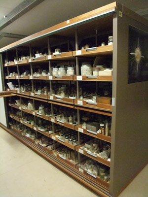
Part of the ocean bottom microfossil residue collection housed in the Department of Palaeontology.


The team have borrowed some of our most significant microfossil residues to test their system and develop future applications. These residues come mainly from the H.M.S. Challenger Collection. The Challenger Expedition of 1872-1876 was the first oceanographic voyage. Prior to this, the ocean bottom was completely unexplored so these residues represent a 'first ever glimpse' of the bottom of the worlds oceans.


All of the organisms discovered during this first dredging of the ocean bottoms, including the microfossils, were described resulting in many new species being found. The foraminiferal type specimens of many of these species are housed here in the Museum and are frequently consulted by visitors. When a new species is described, a type specimen must be designated and deposited in a recognised museum so that future workers can consult them if neccessary.


_DSC3559_blog.jpg

A type slide from the Challenger Foraminifera Collection. It is 7.5cm long.


As the research is being done in North America, it was topical that we also loaned a residue from the Second Albatross Cruise of 1882. The Albatross was reportedly one of the the first vessels built for oceanographic studies and made its maiden voyage from Washington.


Dil Joseph's team in Edmonton have already published on computer aided recognition of foraminifera. It will be very interesting to see how historically significant materials from our collections play a part in future research on this subject. Work on Virtual Reflected Light Microscopy certainly looks to revolutionise how data on microfossil collections will be shared and how we make our collections available in the future.


Special thanks to Lil Stevens who processed this loan. She has now joined the full time curatorial staff and is helping out with micropalaeontology curation.

1

The permanent Dinosaur gallery at the Museum is open every day of the year (except 24 to 26 December) but the recent temporary exhibition 'Age of the Dinosaur' closed last week and now sets off on its travels to another temporary location. Keen eyed visitors to Age of the Dinosaur may have seen some microfossil pictures lurking at the back of the exhibit. Microfossils and dinosaurs are at different ends of the size scale so how can work on microfossils be related to dinosaur research?


DSCF1170_blog.jpgDSCF1169_blog.jpg

The display in the 'Age of the Dinosaur' exhibit including the microfossil images


A neat piece of work published in 2008 by several former colleagues in the Journal Cretaceous Research described some ostracods, spores and pollen from a dinosaur excavation site at Ockley in Surrey. These microfossils show that an Iguanodon died and was buried in a temporary freshwater pond, while the spores and pollen indicate that the Iguanodon lived in a world dominated by certain types of ferns and conifers. The microfossil assemblage tells us that the Iguanodon died in the early part of the Barremian, a stage of the Cretaceous 121-127 million years ago.


Dinosaur officionados will correct me and say that Iguanodon is now called Mantellisaurus. Details about dinosaurs can be found on the Museum web site if you are interested to read further.


In 2001 a large party from the Department of Palaeontology spent two weeks excavating a site at Ockley where the partial skeleton of an Iguanodon had been found by a members of a Geologists' Association field excursion. News of the find had spread very quickly to the popular press so we had to take it in turns to stay overnight in a tent at the site so that we could make sure that is wasn't ravaged by opportunist collectors.


Ockley_Iguanodon_blog.jpg

The parts of the Ockley Iguanodon skeleton that were recovered in the excavation.


I remember being asked to wield a pick axe and shovel so that we could clear the overlying rock from the rock bed that the fossil bones had been found in. Meanwhile my vertebrate colleagues were on their hands and knees preparing away the bones that had become exposed on the surface. These were then taken back to the museum still encased in the rock so that they could be further prepared by David Gray in the Palaeontology Conservation Unit.


My former colleague Susanne Feist-Burhardt collected samples for microfossil study from the dinosaur bed and the beds above and below. These were studied for palynology by MSc student Elly Nye and by David Horne and John Whittaker for ostracod microfossils.


Spores_Pollen_2.jpg

Examples of some of the spores and pollen obtained from the dinosaur site. The palynological record suggests that the dinosaur came to rest on a warm subtropical flood plain surrounded by ferns and conifers.


Scenedesmus2-PH-2_blog.jpg

The freshwater green algae Scenedesmus novilunaris found in the dinosaur bed suggests that its final resting place was a freshwater pond.


Cypridea_blog.jpg

The ostracod Cypridea clavata along with other evidence suggests that the pond dried out periodically.


Samples close to the dinosaur bed produced the first recognisable ostracods from the Ockley site allowing a firm date of the age of the sediments to be established. The single species of the genus Cypridea found suggests that the pond dried out on a regular basis. The eggs that this group of ostracods produces are resistant to dry periods. As a result, their relatives are often found in modern day temporary ponds. More common Cretaceous ostracods that require permanent water bodies are absent from the Ockley assemblage giving further evidence for the temporary nature of the pond where the Iguanodon came to rest.


This was a very interesting project to play a small part in and shows the power of tackling projects as a group. It also shows some excellent potential applications of micropalaeontology so I was very glad that it became part of the 'Age of the Dinosaur' exhibit.

0

Two upcoming events will enable you to see the original specimens and the scale models of the ostracod that showed evidence of sexual reproduction through the use of giant sperm 140 million years ago.


At Science Uncovered on 23 September (see flyer below for details), I'll be on the Palaeontology table from 16.00-17.00. And, a few days before, at 14.30 on 19 September I'll also be taking part in the 'Microscopic sex' talk for Nature Live in the Attenborough Studio in the Darwin Centre.


I hope to see you at one or the other (or both!).


Science Uncovered_E-INVITE.JPG

Giles Miller

Giles Miller

Member since: Apr 21, 2010

This is Giles Miller's Curator of Micropalaeontology blog. I make the Museum micropalaeontology collections available to visitors from all over the world, publish articles on the collections, give public talks and occasionally make collections myself.

View Giles Miller's profile