Skip navigation

The NaturePlus Forums will be offline from mid August 2018. The content has been saved and it will always be possible to see and refer to archived posts, but not to post new items. This decision has been made in light of technical problems with the forum, which cannot be fixed or upgraded.

We'd like to take this opportunity to thank everyone who has contributed to the very great success of the forums and to the community spirit there. We plan to create new community features and services in the future so please watch this space for developments in this area. In the meantime if you have any questions then please email:

Fossil enquiries: esid@nhm.ac.uk
Life Sciences & Mineralogy enquiries: bug@nhm.ac.uk
Commercial enquiries: ias1@nhm.ac.uk

Curator of Micropalaeontology's blog

3 Posts tagged with the coccolith tag
0

My diary this week is illustrated by an item from behind the scenes at the Museum for each day. The week's events include:

  • an offer of an important historical collection for sale
  • work towards digitising an amazing collection of marine plankton pictures
  • hosting an old friend from Brazil
  • a recent donation appearing in a publication
  • preparation for a session on the scanning electron microscope


Monday

Below is an example from our collection of a slide made by the famous slide mounter Charles Elcock in 1880. Slides made by Elcock fetch as much as £350 on the open market so it was very exciting to be approached by a dealer to see if we wanted to acquire his foraminiferal collection and archive. Unfortunately the asking price was significantly more than we can afford.

Elcock_DSC3569.jpgA slide from our collections made by Charles Elcock in 1880.


We regularly buy specimens, and in fact some of my colleagues recently purchased specimens at the Munich Rock and Fossil Show. As far as I know, the last item bought for the micropalaeontology collection was a conodont animal back in the late 1980s.


At the very least this collection offer makes me realise the monetary value placed on historical items in the collection that I look after.


Tuesday

Below is a scanning electron microscope image of a coccolith, part of a collection of over 6,000 images taken by my former colleague Dr Jeremy Young and his collaborators.
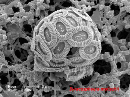
207-23-syra. molischii_SU_blog.jpgScanning electron microscope image of the coccolith Syracosphaera molischii.


Recently graduated micropalaeontology Masters student Kelly Smith is visiting today to help us work up the data on this collection so that we can make information and images available via our website. Because their distribution is controlled largely by temperature, coccoliths in ancient sediments have been used to provide details of past climate change.


Coccoliths make their tiny calcareous shells by precipitating calcium carbonate from seawater so their present-day distribution may have been affected by acidification of the oceans relating to the burning of fossil fuels.


Wednesday

In 20 years at the Museum I have met a lot of people from all over the world, some of whom have visited regularly and subsequently become good friends. I first met Prof. Dermeval do Carmo of the University of Brasilia, Brazil, shortly after I arrived at the Museum. We have enjoyed a long collaboration that has included working together on publications, teaching collections management in Brazil, fieldwork and even holidays. He is visiting with a large, empty suitcase to to pick up the last of the scientific papers identified as duplicate by some of our volunteers.


11563_large.jpg

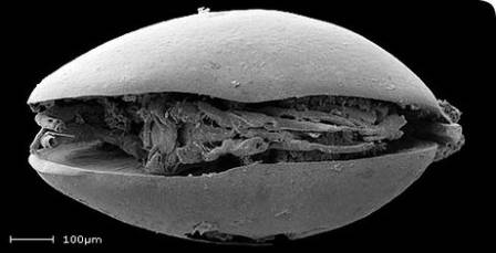
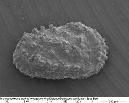
The Brazilian Cretaceous non-marine ostracod Pattersoncypris micropapillosa.


Today's picture is the exceptionally preserved Brazilian Cretaceous non-marine ostracod Pattersoncypris micropapillosa. Dermeval has recently reclassified the genus under the name Harbinia and is on his way to China to look at the type material for this genus. Species related to Pattersoncypris/Harbinia are used to provide information on rock ages and environments of deposition for oil exploration offshore Brazil and W Africa. For more details about their evolutionary significance, see my post on What microfossils tell us about sex in the Cretaceous.


Thursday

Today's scanning electron microscope image is the holotype of a new species of planktonic foraminifera Dentoglobigerina juxtabinaiensis, donated last summer and published last month in the Journal of Foraminiferal Research by University of Leeds PhD student Lyndsey Fox and her supervisor Prof. Bridget Wade.


The Museum collection contains many type specimens that ultimately define the concept for each species. As a result, types are some of our most requested specimens by visitors or for loan.


Fig 7.1c (holotype) u1337a 42xcc_blog.jpg

The holotype of a new species of planktonic foraminifera, Dentoglobigerina juxtabinaiensis.


This species is Miocene in age (roughly 13.5-17 million years old) and was recovered from an International Ocean Discovery Programme core taken from the Pacific Ocean near the equator. Foraminifera, like the coccoliths mentioned earlier, are important indicators of ocean condition and climate, so this unusually well-preserved material is an important contribution to their study. Now the paper has been published I am able to register the details in our database and these will go live on our website in a couple of weeks.


Friday

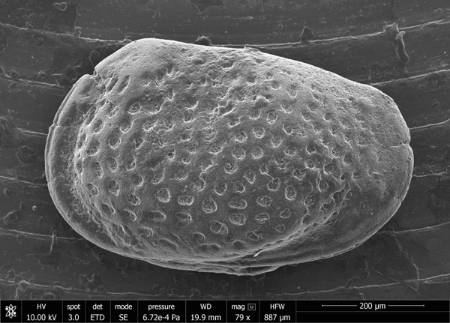
I have a scanning electron microscope session booked next week as I have been approached by a colleague at the British Geological Survey who is preparing a chapter for a field guide on Jurassic ostracods. He would like some images of some of our ostracod type specimens from the Kimmeridge Clay Formation of Dorset.

HULL631_Mandelstamia_maculata_cgmq_107_blog.jpg

The ostracod Mandelstamia maculata from the Jurassic Kimmeridge Clay of Dorset.


Each specimen must be carefully removed from the slides that house them using a fine paint brush and glued to a small aluminium stub about 1cm in diameter. These will be coated in a fine layer of gold-palladium before being placed in the scanning electron microscope chamber next week for photography.


It proves very difficult to identify the relevant specimens as the original publication is pre-scanning electron microscope times and the images taken down a binocular microscope are less than clear. Publishing new, clearer illustrations of each of these type specimens will add considerable scientific value to our collections.


This selection of specimens has hopefully shown the historical, scientific and monetary value of our collections while showing that they are also relevant to important topical issues such as climate change and oil exploration.

0

Every quarter we are required to provide numbers of visitors, enquiries and loans to show how the collections are being used. These are passed to the Museum Trustees and subsequently the Department of Culture, Media and Sport (DCMS) as a performance indicator to help justify the expense of maintaining such an amazing collection.


At the Museum a new loan type - Surrogate Loan - has just appeared on the horizon to record when we send images of our specimens rather than loaning the specimens themselves. Usually I would sigh and say something under my breath about having too much administration. However, as someone who deals with a lot of images of my collection I'm really glad. Recording surrogate loans gives us and the managers who allocate Museum collections management resources a really useful indication of how much the microfossil collection is being used.


Below are a few images I have sent out recently as surrogate loans along with a few comments about them and how they are being used.

cgmx0909_blog.jpg


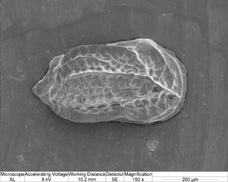
This is the ostracod Lophocythere caesa britannica from the Jurassic (about 160 million years ago) of the UK. The scanning electron microscope picture was taken by me for Prof. Robin Whatley, now retired from University College Aberystwyth, University of Wales. Prof. Whatley has donated a large number of specimens to the Museum during his career and is putting together an extensive publication describing some of them. Robin can't travel easily so his wife Caroline Maybury has been amazingly helpful in preparing the material in our collections for imaging and putting the specimens away afterwards. We would certainly not have sent that many specimens out on loan because they are too fragile and important reference specimens.


cgmx953_blog.jpg


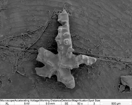
This is an image of the conodont Distomodus staurognathoides from the Silurian (about 425 million years ago) of Iran. It is part of donation by Dr Vachik Hairapetian who has provided us with some very interesting material in recent years. Some of it has been published or like this specimen, is in the process of being published. Conodonts from this area of the world are largely undescribed so it is important that this material is recorded as their occurrences can help with future geological interpretations of the region.


204-59_blog.jpg


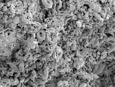
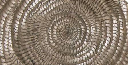
This an extreme close up of a piece of chalk from the Sevens Sisters in Kent taken by my former colleague Jeremy Young. It shows tiny rings of calcareous nannoplankton called coccoliths and was sent to artists Francisco Queimadela and Mariana Calo who visited in February. This followed an open day of the micropalaeontology collections associated with an exhibition at the Gasworks Gallery at the Oval, London. They are also doing an exhibition at the Gasworks Gallery.


cgmx0673_blog.jpg

This is another ostracod, this time from the Coralline Crag of Norfolk donated in 2011 by Dr Adrian Wood of Coventry University. I took scanning electron microscope images of all the specimens in his donation to help with his publications on ostracods from East Anglia. Adrian has been very helpful in preparing the specimens for accession to our collections by labelling slides and providing electronic data that can be easily incorporated into our database.


Throughout my career as a curator I have sent a lot of specimen images to enquirers so I'm very glad that this practise in now being recorded as a performance indicator. Hurrah for surrogate loans!

0

Microfossils of the day

Posted by Giles Miller Aug 12, 2011

To celebrate the United Nation's Year of Biodiversity last year, the Museum published details of a different species every day on its web site under the title Species of the Day. These records were delivered last week to another web site The Encyclopedia of Life. Each species was chosen and written about by a museum scientist so this week's blog is to point you in the direction of the microfossils which were chosen for their importance in studies on climate change, ocean acidification, north sea oil exploration and the fossil record of sexual reproduction. Follow the links below to find out more about each species and the groups to which they belong.


ehux_coccospheres_51005_1.jpg

Emiliania huxleyi


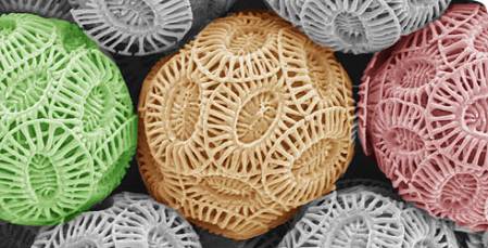
Emiliania huxleyi is a coccolithophore which is a unicellular plant that lives in the upper layers of the ocean and forms tiny calcareous coccolith plates like the ones you can see above. These are artificially coloured images from a scanning electron microscope. This very high powered microscope is needed as they are only tens of microns in size and as a result are usually referred to as nannofossils. The ones above are only slightly larger than a thousanth of a millimetre in size. If you were to dip a bucket in the ocean you could find literally tens of thousands of these types of cells. In early summer, E. huxleyi forms enormous blooms across the northwest European shelf that can be seen from space. Coccoliths are susceptible to changes in climate and ocean acidification. This, combined with an excellent fossil record makes them an essential group in the study of recent changes to our oceans and environment.


11563_large.jpg

Harbinia micropapillosa


Harbinia micropapillosa is an ostracod, a microscopic crustacean with two calcareous shells. Ostracods can be found in virtually any current aquatic environment and very rarely on land in damp habitats near to water. They have an extensive fossil record because their two shells preserve well as fossils but usually the soft body parts decay soon after death. H. micropapillosa is exceptional because the soft body parts have been preserved in a rock formation that is roughly 140 million years ago. Recent analysis using new techniques has shown the reproductive organs of this ancient organism are identical to those of present day ostracods and suggest that they reproduced using giant sperm back in the Cretaceous period. If you can't wait to find our more about this interesting fossil then follow the link above. However, I will be expanding the story of these important specimens in our collections as the subject of a future blog.


29316_large.jpg

Nannoceratopsis gracilis


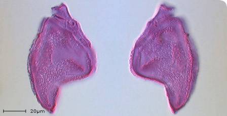
Nannoceratopsis gracilis is a dinoflagellate cyst from the Jurassic period about 145-200 million years ago. Dinoflagellates are marine photosynthetic algae that play an important role at the base of the food chain and the carbon cycle. At stages throughout their life cycle they form resistant organic cysts that can be found in the fossil record by dissolving suitable rocks in nasty acids like hydroflouric acid. Nannoceratopsis is one of the earliest forms of dinoflagellate cyst so studies of this genus can tell us a lot about the early evolution of dinoflagellates. The shape is also very distinctive and easily recognisable. N. gracilis can be found in rocks 168-185 million years old and can therefore be used, on its own or in association with other fossils, to accurately date rocks.

15351_large.jpg

Nummulites gizehensis


I mentioned Nummulites gizehensis is a member of the Foraminifera in my second blog and showed a picture of the pyramids at Gizeh that are constructed from rocks that contain this species. The genus Nummulites is a member of a group called the "Larger Foraminifera" that build multichambered shells up to 15cm in size despite being a single celled amoeba. The chambers like the ones shown above can only be seen by breaking the shells apart or making specially oriented thin sections of the rocks they are found in. Sometimes symbiotic green algae also lived in the chambers, providing products of photosynthesis to the amoebe while using the shell as protection. N. gizehensis lived during the Middle Eocene epoch about 37-48 million years ago, in shallow marine conditions and can be used as a marker to show the age of rocks that contain them, particularly in the oil region of the Middle East.


Finally a big thank you to my former colleagues Jeremy, Susanne and Clive who originally wrote about three of these beautiful microfossil species of the day.


Giles Miller

Giles Miller

Member since: Apr 21, 2010

This is Giles Miller's Curator of Micropalaeontology blog. I make the Museum micropalaeontology collections available to visitors from all over the world, publish articles on the collections, give public talks and occasionally make collections myself.

View Giles Miller's profile