Skip navigation

The NaturePlus Forums will be offline from mid August 2018. The content has been saved and it will always be possible to see and refer to archived posts, but not to post new items. This decision has been made in light of technical problems with the forum, which cannot be fixed or upgraded.

We'd like to take this opportunity to thank everyone who has contributed to the very great success of the forums and to the community spirit there. We plan to create new community features and services in the future so please watch this space for developments in this area. In the meantime if you have any questions then please email:

Fossil enquiries: esid@nhm.ac.uk
Life Sciences & Mineralogy enquiries: bug@nhm.ac.uk
Commercial enquiries: ias1@nhm.ac.uk

0

Curators at the Museum receive donations or offers of donations of new specimens on a regular basis. An interesting rock made almost entirely of microfossils arrived from Brazil a couple of weeks ago. We have not officially accepted this donation yet, but this is an interesting acquisition story about an interesting specimen.


DSCF1112_blog.jpg

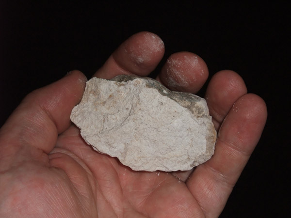
The rock specimen offered for us as a donation


The donor Prof. Dr. José Luiz Lorenz Silva offered the specimen to us following his publication on a rock called a spongillite which was found at Très Logoas in the State of Matto Grosso do Sul, Brazil. This rock was deposited in the Late Pleistocene period about 37,000 years ago in a freshwater pond. The microfossils contained suggest that the climatic conditions were colder at that time and that it was deposited in a slightly acidic peat bog pond.


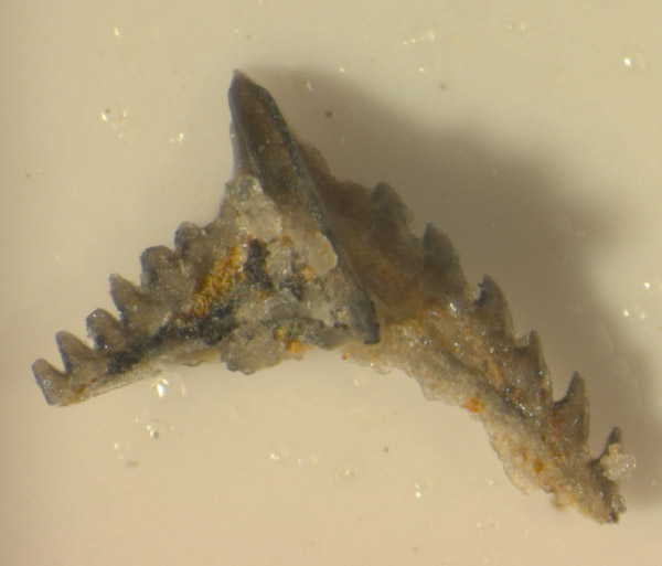
Following the donation, my Erasmus student Angelo Mossoni and I placed a fragment of the rock under the scanning electron microscope to see what it contained. The rock is very light as it is composed almost completely of the siliceous remains of sponges and diatoms. Diatoms are algae that secrete a characteristic shell wall or frustrule of silica. They can be found in the fossil record as far back as the Jurassic period and are commonly used in the present day as indicators of water quality.


cgmx0648_blog.jpg

A scanning electron microscope image of part of the rock showing the remains of thousands of diatoms


At the start I mentioned that we had not officially acquired this specimen. All future donations are carefully scrutinised so that we are sure that the material has been donated to us without contravening laws of the country that it was collected from. Brazilian law states that specimens cannot be collected from their country and sent abroad unless relevant agreements exist between the institutions in Brazil and the institutions accepting the donations.


To account for this, we have registered the specimen as "Object Entry". This means that we are not certain just yet if we have the correct paperwork and agreements in place to accept it and acknowledge that we do not officially hold title to its ownership. Specimens sent to us for identification or specimens belonging to other institutions but being studied here are commonly registered here as "Object Entry" until they are returned to their owners.


cgmx0649_blog.jpg

Some more diatoms under the scanning electron microscope. The scale on the picture is 10 microns which is 0.001 of a millimetre.


As you can see from the scanning electron microscope images above, it is certainly a very interesting rock. Museum policy on acquisition states that we will seek to acquire specimens if one of the following is satisfied: it helps to bridge or fill gaps in collections, it fulfills a research need, it complements existing specimens in our collection, it has potential for public education or display, it is scientifically or historically significant.


The spongillite certainly covers several of these criteria and may well be of interest to the research of scientists in the Botany Department studying diatoms. My colleague Martha Richter is currently in Brazil and will be checking if the documentation we have is acceptable. Until we are sure, this very interesting rock specimen won't officially be making its way into our collections just yet.

0

Microfossils of the day

Posted by Giles Miller Aug 12, 2011

To celebrate the United Nation's Year of Biodiversity last year, the Museum published details of a different species every day on its web site under the title Species of the Day. These records were delivered last week to another web site The Encyclopedia of Life. Each species was chosen and written about by a museum scientist so this week's blog is to point you in the direction of the microfossils which were chosen for their importance in studies on climate change, ocean acidification, north sea oil exploration and the fossil record of sexual reproduction. Follow the links below to find out more about each species and the groups to which they belong.


ehux_coccospheres_51005_1.jpg

Emiliania huxleyi


Emiliania huxleyi is a coccolithophore which is a unicellular plant that lives in the upper layers of the ocean and forms tiny calcareous coccolith plates like the ones you can see above. These are artificially coloured images from a scanning electron microscope. This very high powered microscope is needed as they are only tens of microns in size and as a result are usually referred to as nannofossils. The ones above are only slightly larger than a thousanth of a millimetre in size. If you were to dip a bucket in the ocean you could find literally tens of thousands of these types of cells. In early summer, E. huxleyi forms enormous blooms across the northwest European shelf that can be seen from space. Coccoliths are susceptible to changes in climate and ocean acidification. This, combined with an excellent fossil record makes them an essential group in the study of recent changes to our oceans and environment.


11563_large.jpg

Harbinia micropapillosa


Harbinia micropapillosa is an ostracod, a microscopic crustacean with two calcareous shells. Ostracods can be found in virtually any current aquatic environment and very rarely on land in damp habitats near to water. They have an extensive fossil record because their two shells preserve well as fossils but usually the soft body parts decay soon after death. H. micropapillosa is exceptional because the soft body parts have been preserved in a rock formation that is roughly 140 million years ago. Recent analysis using new techniques has shown the reproductive organs of this ancient organism are identical to those of present day ostracods and suggest that they reproduced using giant sperm back in the Cretaceous period. If you can't wait to find our more about this interesting fossil then follow the link above. However, I will be expanding the story of these important specimens in our collections as the subject of a future blog.


29316_large.jpg

Nannoceratopsis gracilis


Nannoceratopsis gracilis is a dinoflagellate cyst from the Jurassic period about 145-200 million years ago. Dinoflagellates are marine photosynthetic algae that play an important role at the base of the food chain and the carbon cycle. At stages throughout their life cycle they form resistant organic cysts that can be found in the fossil record by dissolving suitable rocks in nasty acids like hydroflouric acid. Nannoceratopsis is one of the earliest forms of dinoflagellate cyst so studies of this genus can tell us a lot about the early evolution of dinoflagellates. The shape is also very distinctive and easily recognisable. N. gracilis can be found in rocks 168-185 million years old and can therefore be used, on its own or in association with other fossils, to accurately date rocks.

15351_large.jpg

Nummulites gizehensis


I mentioned Nummulites gizehensis is a member of the Foraminifera in my second blog and showed a picture of the pyramids at Gizeh that are constructed from rocks that contain this species. The genus Nummulites is a member of a group called the "Larger Foraminifera" that build multichambered shells up to 15cm in size despite being a single celled amoeba. The chambers like the ones shown above can only be seen by breaking the shells apart or making specially oriented thin sections of the rocks they are found in. Sometimes symbiotic green algae also lived in the chambers, providing products of photosynthesis to the amoebe while using the shell as protection. N. gizehensis lived during the Middle Eocene epoch about 37-48 million years ago, in shallow marine conditions and can be used as a marker to show the age of rocks that contain them, particularly in the oil region of the Middle East.


Finally a big thank you to my former colleagues Jeremy, Susanne and Clive who originally wrote about three of these beautiful microfossil species of the day.

1

The Erasmus Programme is European Union funded and enables higher education students in 31 European countries to study for part of their degree in another country. This is exactly what Italian Geology student Angelo Mossoni is doing here at the Museum this summer as part of his masters degree at Cagliari University.


DSCF1035_blog.jpg

Angelo working in the Micropalaeontology Laboratory


Angelo contacted me in early 2011 asking if I had any projects that I needed doing on conodont microfossils. Conodonts are the teeth of an extinct worm-like organism that are used widely for investigating the age of rocks between 200-500 million years old. This sort of information is very useful for oil or mineral exploration companies as well as for revealing the geological history of rock formations.


Finding conodonts can be a long process. The first stage is to dissolve in vinegar, limestones or other rocks that were deposited in shallow to deep seas. This produces large residues of microscopic fragments less than a millimetre in size that need to be examined under a microscope. I had already dissolved tens of kilogrammes of limestones from Oman so Angelo's first task was to help examine the residues.


Angelo has previous experience of this type of work but has not used separating techniques to reduce the size of the residue needing to be examined. He was introduced to a method using a heavy liquid sodium polytungstate that concentrates the heavier fractions of the residues that include the conodont microfossils. This means that less time is needed to examine the results under a microscope, a process that can sometimes take weeks or even months.


DSCF1039_blog.jpg

Adding residues to the heavy liquid sodium polytungstate


The heavy fraction then needs to be examined under the microscope and a fine paint brush used to transfer the conodonts into a separate cavity slide for further examination. Angelo and I then worked together to choose which specimens should be illustrated. Angelo was then taught to use the Axiocam Imaging System to take good publication quality colour images of the best specimens that he found (see below).


17a montage_blog.jpg

One of the conodont specimens found by Angelo. It is just over a millimetre in length.


The very best specimens were also illustrated using a scanning electron microscope so that the conodonts we found could be classified and used to provide a geological age for the sample. Precise dating of rocks from Oman is potentially of interest to oil companies in this region. Angelo found many other interesting fragments of fish and microfossils that will be published along with the conodonts in the future.


DSCF1045_blog.jpg

Studying some microscopic fish remains on the scanning electron microscope


Part of the strategic plan for the Museum is to 'provide a unique and personalised experience for learners, through engagement with real science, scientists and specimens'. We have certainly done this by teaching Angelo new techniques in the study of conodonts while he helps with a research project that has significantly enhanced our collections. More importantly we have enabled Angelo to achieve some of the goals of the Erasmus Programme by experiencing study in a laboratory away from is own country and greatly improving his English.


Angelo leaves at the end of August but that will not be the end of his association with the Museum. During his stay he has found many interesting new specimens that we will eventually publish together. Hopefully this will also be a great help to him in his future career as a micropalaeontologist. He has certainly done a large amount of very useful work during his time here for which I am very grateful.

Giles Miller

Giles Miller

Member since: Apr 21, 2010

This is Giles Miller's Curator of Micropalaeontology blog. I make the Museum micropalaeontology collections available to visitors from all over the world, publish articles on the collections, give public talks and occasionally make collections myself.

View Giles Miller's profile