Skip navigation

The NaturePlus Forums will be offline from mid August 2018. The content has been saved and it will always be possible to see and refer to archived posts, but not to post new items. This decision has been made in light of technical problems with the forum, which cannot be fixed or upgraded.

We'd like to take this opportunity to thank everyone who has contributed to the very great success of the forums and to the community spirit there. We plan to create new community features and services in the future so please watch this space for developments in this area. In the meantime if you have any questions then please email:

Fossil enquiries: esid@nhm.ac.uk
Life Sciences & Mineralogy enquiries: bug@nhm.ac.uk
Commercial enquiries: ias1@nhm.ac.uk

5

This week a surplus set of plaster microfossil models were transferred to the Department of Geology, University Leicester UK to help with teaching micropalaeontology to undergraduate students. The two sets of models were made by 19th Century scientists d'Orbigny (1802-1857) and Reuss (1811-1873), who were some of the very earliest micropalaeontologists.

DSCF1265_blog.jpg


A set of d'Orbigny models from the Museum collections. Some look a dirty brown colour but in fact this is an original feature to show the difference between models based on modern species (white) and fossil ones (brown). This set was previously mounted for display in the Museum galleries.


DSCF1273_blog.jpg


A drawer of d'Orbigny models of Foraminifera. (Skaters on the Museum ice rink can be seen in the background)


The famous French scientist Alcide d'Orbigny quickly recognised the difficulty in portraying his work on microfossils to a wider audience because of the small size of the specimens. He carved scale models of foraminferal microfossils from limestone and these originals are in the Museum national d'Histoire naturelle in Paris. He used these to create plaster replicas that he sold in sets to accompany his publication of the first classification of the Foraminifera which was first published back in 1821.


DSCF1281_blog.jpg

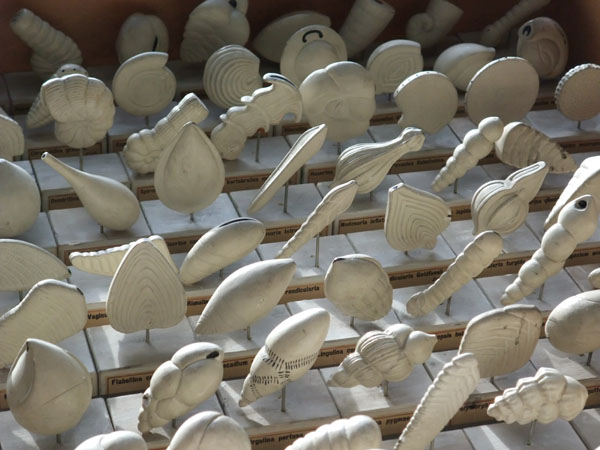
A 'Plaster Army' of Reuss & Fric models arranged in rows reminiscent of the 'Terracotta Army'.


The second set was made by Vaclav Fric (1839-1916) under the supervision of Anton Reuss who was similarly looking to illustrate his classification of the Foraminifera. For more information about these models and other microfossil models at the Museum there is a publication in the Geological Curator. A paper was recently submitted for publication in a Special Publication of the Geological Society as a contribution to a set of papers on the history of study of the Foraminifera.


DSCF1285_blog.jpg

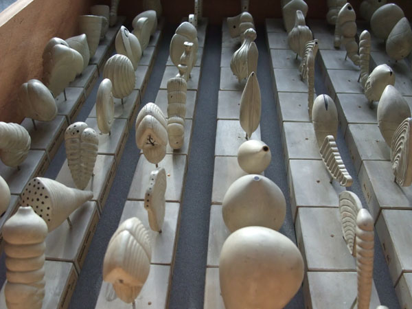
Some more Reuss & Fric models. The black spots show the openings or 'foramen' common to and therefore giving rise to the name 'Foraminifera'.


If these models are so important, why are we letting them go from the Museum? Firstly we already have three registered sets of these models in our collections (some are illustrated above). One of these three sets is is on the salvage list for the Palaeontology Department. This means that these will be some of the first items to be saved from the building should there be some sort of disaster and it is deemed safe to do so.


Secondly, the model sets on their way to the University of Leicester were never formally accessioned into the Museum collections so we are able to send them on without having to deaccession them. They are slightly worn as they have previously been used for teaching micropalaeontology to postgraduate students. Currently there are limited opportunities for postgraduate study of micropalaeontology so it is very good to know that a new course is starting at the University of Birmingham in September 2012.


I would argue that sending these models to a university to help inspire a new generation of micropalaeontologists is exactly the sort of use that d'Orbigny and Reuss would have wanted for their models rather than for them to sit in a box in a dusty corner of my office...

1

Some of the Museum's most important ostracod specimens were re-examined recently using synchrotron technology. The results published in the journal Science showed that these very delicate but exquisitely preserved fossils gave evidence for reproduction using giant sperm back in the Cretaceous period about 100 million years ago.


11563_large.jpg


A scanning electron microscope of an exceptionally preserved ostracod from Brazil showing details of unusually preserved soft body parts.


From images obtained by scanning electron microscope we have known since the 1970s that the Cretaceous ostracod Harbinia micropapillosa was almost identical in body form to modern day examples. Usually ostracods, microscopic crustaceans that inhabit aquatic environments, leave only their calcareous shells in the fossil record. However, these exceptional fossils from Brazil include details of their organic soft body parts not normally fossilised.


These specimens were first found by legendary evolutionary biologist Dr Colin Patterson while he was studying the fossil fish from the same rock formation. He passed them to Dr Ray Bate who published them under the name Pattersoncypris. However, some ostracod workers now believe that they should be classified under the name Harbinia which was first described by a Chinese worker in 1959 and therefore takes naming priority.


Grenoble 125_blog.jpg

The European Synchrotron Research Facility (ESRF) at Grenoble in France.


In 2007 we had a request by Dr Renate Matzke-Karasz (University of Munich) and a group of co-workers to take our specimens to Grenoble in France to have them analysed in the synchrotron beam ID19. A synchrotron is a giant ring where electrons are accelerated to great speeds and then bent into a circular path by magnets. Strong magents are used which cause the electron beam to deviate and at this point a very bright, intense synchrotron x-ray is emitted. Sometimes synchrotrons are referred to as diamond light sources as a result. These very intense synchrotron x-rays are then focussed into a beam which can be used for analysis at a sub micrometer scale ideal for our microfossils.


Some types of modern day ostracods are well known for their use of giant sperm in reproduction. Dr Matzke-Karasz and her co-workers were interested to see if our fossil specimens (Robin Smith thesis collection) contained any evidence for giant sperm or the organs responsible for its production and storage. As the curator of the specimens it was my job to transport them safely to Grenoble and to handle them while they were being analysed. I also took part in the analysis which went on all day and all night for two days. Fortunately we did get some sleep as there were four of us. We took it in turns with two of us analysing the fossils and two analysing the comparative modern specimens in 6 hour shifts.


Grenoble 063_blog.jpg


Positioning the specimen so that it is aligned with the beam. (Don't worry about the scary red lines. The beam was only switched on when we were all safely out of the room!).


Grenoble 038_blog.jpg

Dr Radka Symonova (then at Charles University, Czech Republic), Renate, Dr Paul Tafforeau (ESRF) and Dr Robin Smith (Lake Biwa Museum, Japan) examining some early scans in our experimental cabin home for the two days.


The specimens were placed in the beam and then rotated 180 degrees while 1500 x-ray cross sections were taken at regular intervals. These x-ray images were then combined together using specially designed software to produce 3-dimensional images (Holotomographic reconstructions). Although we could immediately see evidence for important internal structures while we were analysing the specimens, a lot of work was still required to produce the final results. The slices that make these 3-dimensional images were analysed for internal structures by Renate and her team back in Germany. Artificial colours were painstakingly added to each slice by hand to show these structures more clearly.


Grenoble 054_blog.jpg


One of the x-ray cross sections of a fossil specimen before it was combined into a 3-dimensional image.


The results clearly showed differences between males and females. The males had distinctive tubes in the position where modern day ostracods have  a sperm pump called a Zenker's Organ. The females had inflated sacks in the position where modern day ostracods have sperm receptacles. These are only inflated once they have been impregnated with giant sperm. Our results had shown that this reproductive strategy had been in place more than 100 million years ago.


Video of a female specimen of Harbinia micropapillosa. The orange sacks are the sperm receptacles.


So why is this important? As I showed in the dinosaur exhibition blog item, it is vital to know how organisms reproduce so that you can correctly interpret their fossil record and distribution in modern day environments. Ostracods are often restricted to particular environments and can be useful indicators of changes in climate. This particular ostracod species is common in Cretaceous non-marine sediments offshore Brazil and is therefore of interest to oil exploration companies as a marker for key rock formations.


Reproduction with giant sperm is not just restricted to the ostracods as other organisms including fruit flies and some types of frog also use this strategy. The evolutionary significance and history of this type of reproductive strategy is still unclear. What is certain is that specimens in the Museum collections show that this was also happening over 100 million years ago!


DSCF1135_blog.jpg


Some acrylic palm of the hand sized scale models produced from the 3-D synchrotron scans and used at the "Science Uncovered" event.


Giles Miller

Giles Miller

Member since: Apr 21, 2010

This is Giles Miller's Curator of Micropalaeontology blog. I make the Museum micropalaeontology collections available to visitors from all over the world, publish articles on the collections, give public talks and occasionally make collections myself.

View Giles Miller's profile