Skip navigation

The NaturePlus Forums will be offline from mid August 2018. The content has been saved and it will always be possible to see and refer to archived posts, but not to post new items. This decision has been made in light of technical problems with the forum, which cannot be fixed or upgraded.

We'd like to take this opportunity to thank everyone who has contributed to the very great success of the forums and to the community spirit there. We plan to create new community features and services in the future so please watch this space for developments in this area. In the meantime if you have any questions then please email:

Fossil enquiries: esid@nhm.ac.uk
Life Sciences & Mineralogy enquiries: bug@nhm.ac.uk
Commercial enquiries: ias1@nhm.ac.uk

Citizen science blog

11 Posts tagged with the dna tag
1

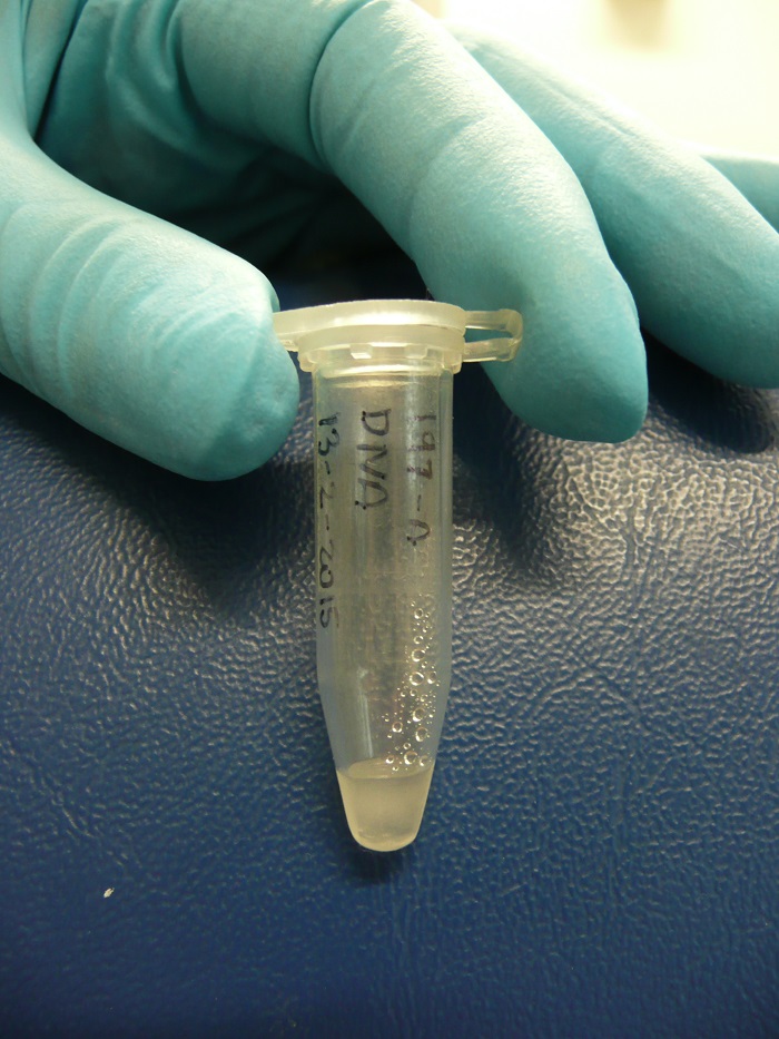
Today one of our Microverse citizen science project participants, Robert Milne, presents his own interpretation of the results of the microbial samples collected from Mid Kent College in Gillingham where he is a student:


The results:


Despite our best efforts, the samples we obtained for the Microverse project were taken in different weather conditions, at slightly different times, in slightly different areas of the building, and all three samples were taken from walls facing different directions. The materials of the surfaces we sampled were brick, glass and metal.


206A_JPG0001.jpg

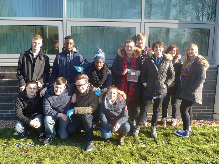
Mid Kent College building, swabbed by The Microverse participants.


From the results below, it can be seen that all three surfaces have about the same number of OTUs, (Operational Taxonomic Units, a phrase to indicate taxonomic groupings in microorganisms), but this does not mean that each surface has the same number of individual microorganisms. The number of genetic sequences varies greatly.


Sample Area A

(brick)

Sample Area B

(glass)

Sample Area C

(metal)

Number of genetic sequences generated88,264120,49827,894
Number of OTUs2,1982,1071,960
% of sequences that were from Archaea0.02%0.00%0.00%
% of sequences that were from Bacteria75.62%88.76%87.75%
% of sequences that were from Eukaryotes24.36%11.24%12.19%


Table 1: Results from samples of microorganisms swabbed from brick, glass and metal, at Mid Kent College, Gillingham, (% rounded to 2 decimal places).


The glass surface has generated the most genetic sequences while metal has generated the least. This could mean that the bacteria on the surface of the glass are more successful than the ones on the metal, for instance.


206A_JPG0002.jpg

Sample Area A - Brick.


The image above shows the brick wall from which the first sample was taken. This wall had the most eukaryotic cells present, in which the majority of them contained chloroplasts (these are the organelles of plants that convert light energy into sugar).


This wall faces southwest and a wall facing south of any kind will always receive the most sunlight on it during the day, which could explain the increased chloroplast numbers compared to the other two surface areas we sampled. The fact that this wall was also close to a lot of grass could also play a part in these numbers.


206B_JPG0002.jpg

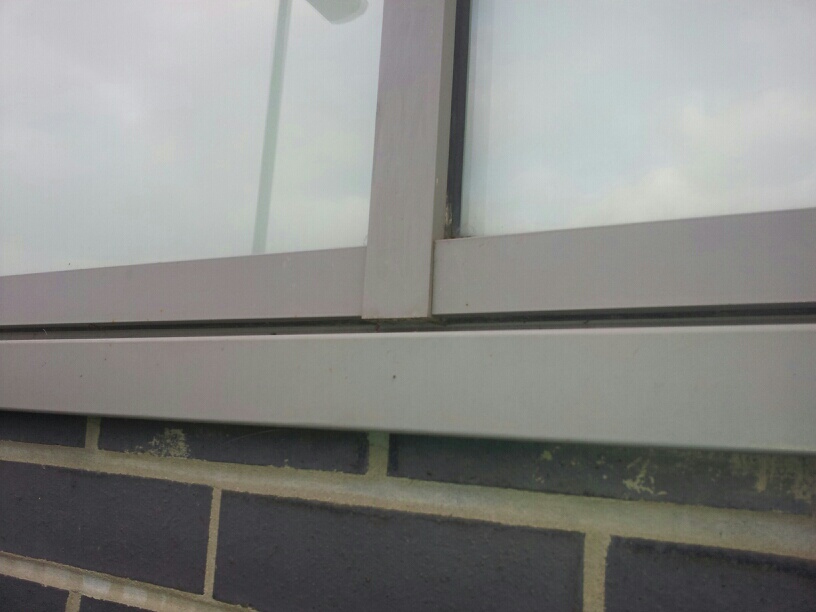
Sample Area B - Glass.


The image above shows the second surface sampled, which was glass. This had the most genetic sequences found out of all three of the surfaces we swabbed. There were, however, less eukaryotic cells on the glass and metal surfaces than on the wall.


This could be because the smooth surface of the metal and the glass meant that less eukaryote cells could remain on the surfaces for prolonged periods. The eukaryotic cells (represented by the mitochondria and chloroplast sequences in the sample) could have originated from natural wildlife around the area, such as a snail's trail or some spider webbing.


206C_JPG0002.jpg

Sample Area C - Metal.


Most of the eukaryote sequences found in all samples were chloroplasts, rather than mitochondria. This probably means the surfaces always have some form of sunlight on them, which is somewhat true since all the surfaces faced either west or east to some extent.


206 Relative abundance chart.jpg

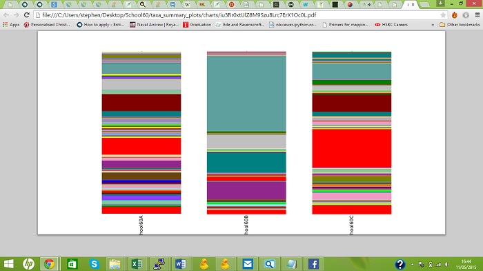
Figure 1: The relative abundance of bacterial phyla, archaea, mitochondria and chloroplasts in the three samples.


Possible uses:


One of the prime examples for undertaking this feat of exploring more of the microbiological world is the need to find better antibiotics; resistance to antibiotics is an increasing threat in the world of medicine. Antibiotic discovery can occur via the identification of bacteria that produce chemical substances that kill or inhibit the growth of other bacteria. Once identified the chemical substance can potentially be cultured and used as a treatment to kill off bacterial infections.


Exploring the countless surfaces outside in the world is a treasure trove of information that could lead to the discoveries of new bacteria that can be used effectively as a source for an antibiotic.


However, it can also be considered that a new resilient bacteria could be discovered that can survive without much water for a long time, which may, just maybe, hold a specific DNA sequence to help relieve the effects of hunger and thirst in patients that must undergo a fast before an operation (such as colon screening). It can open up a number of new doors to the world of medicine, and with a huge percent of areas still not investigated, it could only be a matter of time before huge changes are discovered.


Robert Milne


Thank you Robert! Robert Milne is a student of Mid Kent College, who has just finished his second year of an Applied Science Level 3 course. He has a keen interest in biochemistry and genetics and hopes to enrol this Autumn on an Undergraduate degree in Chemistry at the University of Greenwich. To find out more about the Museum's citizen science projects, see our website.

0

This week Dr. Della Hopkins tells us about how the Decoding Nature project takes school students out on field trips and involves them in the Museum's science research.


In June, a group of ‘scientists in the making’ from Pimlico Academy joined up with a small band of research scientists from the Museum as part of a long running project called Decoding Nature. Decoding Nature is a Museum-run venture which delivers residential science courses to school children aged 8-18.


The courses take place at The Old Malthouse School near Wareham in Dorset, and combine learning with original, ongoing scientific research. Over the years the project has evolved and included a wide range of scientists with varied areas of expertise. Each course is different, ensuring that the children are taking part in cutting edge research that will be used for publication.


Image1 CocoPimlico.JPG

Amazing lichen communities on Dorset trees - a winning photo from the photography competition. Image credit: Coco from Pimlico.


For this particular course our budding scientists from Pimlico Academy were set several tasks, to aid renowned Lichenologist Holger Thues with several important research questions. The week began with an introduction to lichens and a recap on classification and how to use keys, before launching into investigations into the species composition of spice lichens; bags of mixed lichen species sold as cooking ingredients in many Asian supermarkets.


Image 2 Crookshank OMH.jpg

Recording the lichens on coastal trees. Image Credit: Annabel Crookshank, The Old Malthouse.


Next we took to the fields to carry out an air quality survey (designed by the OPAL project), using nine target lichen species growing on tree branches to give an indication of pollution levels. Three of these lichens are very sensitive to pollution, three are ‘intermediate’ and the final three are nitrogen-tolerant. The children’s findings were of great interest, showing that the presence of sheep dramatically altered the lichen community, with the nitrogen sensitive species nowhere to be found. Less than a mile away, by the coast, these species were flourishing.


Image 3 Crookshank OMH.jpg

Comparing the lichen species on trees in grazed fields. Image credit: Annabel Crookshank, The Old Malthouse.


Once they had honed their lichen-identification skills we headed out to Dancing Ledge, a beautiful area on the coast where the cliffs meet the sea. The students carried out a series of transects to answer questions about the habitat preferences of the coastal lichens, such as whether the rocks were sea- or land-facing, vertical or horizontal, and how close to the sea they were. We made some surprising discoveries, and were able to make some collections to take back to the Natural History Museum.


Image 4 Hopkins NHM.JPG

Making a (permitted!) lichen collection at Dancing Ledge.


Finally we used DNA barcoding techniques on a number of European and British samples of the lichen Verrucaria pachyderma. This involved working in the laboratory to extract DNA, and amplify it to a level which could be taken to the museum for the sequences to be read. This was very important work, and great care was needed with the samples. Everyone managed the task superbly, and we were successful in extracting the DNA.


Image 5 Hopkins NHM.jpg

Ready and prepared for DNA extraction.


Once we left Dorset, and returned to the museum, the DNA samples were sequenced. Genetic analysis of the lichens collected from Europe show that the British collections may in fact represent an entirely separate species. The next step is to expand the collection range and analyse more genetic markers, but the results so far are already a big surprise and wonderfully exciting.


Image 6 Crookshank OMH.jpg

Holger Thues explaining the different habitat preferences of the lichens. Image credit: Annabel Crookshank, The Old Malthouse.


The week was full of games to encourage learning, especially with the more tricky subjects such as DNA barcoding. The children entered each challenge with enthusiasm, whether they were in the lab extracting DNA, or buzzing around the field as worker bees collecting their sand pollen. We also found time to learn about other techniques used by scientists in botany (plants) and entomology (invertebrates).


To find out more, visit our Decoding Nature webpage.

You can also find out how to participate in OPAL surveys here.


Della Hopkins


Dr Della Hopkins has worked at the NHM for 6 years, and has managed the Decoding Nature project for the past 5. She previously worked on seed conservation with the Millennium Seed Bank at Kew. She studied Environmental Biology at Royal Holloway University of London, followed by an MSc in Ecology (Bangor) and her PhD from London investigated heathland conservation and restoration.


Della runs the Decoding Nature project with Dr Robert Dyer (molecular laboratory assistant).

0

Citizen Science Project Manager Lucy Robinson introduces a Q&A with Dr. Anne Jungblut.


In an earlier blog post, a group of students from Bedford Girls’ School described their recent visit to the Museum. The girls had taken part in The Microverse, collecting samples of microorganisms from buildings and sending them to the Museum for DNA analysis, and were keen to meet the scientists involved to find out more. We arranged for them to meet the lead researcher on the project, Dr. Anne Jungblut, to ask her some questions about the project and her wider research. We thought you might like to hear her responses:


Q. What inspired you to set up this project?


A. Of all the life on Earth, only a relatively small proportion are the plants, animals and fungi that we can see – the vast majority are microscopic. My research takes me all over the world, where I collect samples of microorganisms and study them using DNA technologies to better understand these important organisms. I’ve done a lot of work in the Antarctic, but I thought to myself that it would be really cool to also look at the microorganisms in the UK, in particular on buildings. There’s been very little research into the microorganisms that live on buildings in towns and cities to see what role they are playing in urban ecosystems. So I contacted Lucy and Jade in the Museum’s citizen science team as this research would require lots of samples to be collected across the country and I thought citizen science – collaborating with members of the public – could be a good option. Together we developed The Microverse project.


TheMicroverse_Green.jpg


Q. What are you looking for in the data – what kind of patterns?


A. Firstly, I’m looking for the overall diversity of microorganisms. They are such an understudied group that these data will give us a baseline understanding of microorganism diversity on buildings. I’m also looking for differences between building materials – we asked participants to sample three different building materials so we will have a lot of different materials to compare.


We also asked you to record a number of different variables that might affect diversity for example the distance to the nearest road and the nearest vegetation. These variables show us possible pollution levels, or semi-natural habitats that microorganism may have colonised the wall from. I’m interested to see what influence the proximity to roads and vegetation/soil has on the microbial diversity.


I’m also keen to see whether unique locations have different communities of microorganisms. Some sample sites are quite unusual e.g. on land contaminated by heavy metals, and on a pier over the sea. Will these buildings have very different communities of microorganisms to the other samples?


This research will also allow us to formulate more detailed hypotheses and refine our research questions. We are also inviting participants to suggest new hypotheses and future directions for the research. Ideas can be emailed to microverse@nhm.ac.uk.


Untitled-1.jpg

Arachnula impatiens, a microorganism found on walls, is a predatory protozoan


Q. How will the Museum judge how accurate the data are?


A. The schools and community groups taking part in The Microverse are carrying out exactly the same method to collect samples as a professional Research Assistant would have done. This means that samples need to be collected under sterile conditions, following a strict protocol.

When we were developing the project, we chose A-level students (or equivalent) as the main audience as they’re committed to science, and we felt they would be more likely to carry out the survey correctly and understand the importance of sterile working compared to other potential audiences we considered e.g. primary school students. Collecting samples in the right way is the first step to ensuring data accuracy.


Once we receive the samples, there are a number of ways we can check the accuracy of the data. After the PCR step, gel electrophoresis checks whether enough genetic material is present in the sample. The sequencing process also removes low quality sequences (ones that are too short in length) which will not give reliable results. The great thing about using DNA technologies for identification is that it’s very accurate and doesn’t rely upon human ability to make a correct identification.


Participants record details about their building surface, but we also ask them to send us photographs, so we can double check if we are unsure about the accuracy of a piece of information, or if it’s an unusual building surface that we need to be able to see to properly interpret the results.

Finally, when we sequence the data, the output shows us how many mitochondria sequences were generated which indicates how much animal DNA there was in the sample. If a sample had been contaminated e.g. by someone’s hands touching the swab, it would show up as a very high number of mitochondria and we would be able to exclude that sample from our analyses. Luckily this hasn’t yet happened.


TrinityCatholicSchool226A.jpg

Students from Trinity Catholic School collecting samples for The Microverse project


Q. Have you seen any microbes in The Microverse samples that you haven’t seen before?


A. Not yet. Samples are still coming in and are being sequenced so we only have very early results from a few sample sites. I will know more when all the samples have been sequenced and analysed. The sequencing we are doing is not always able to identify a microorganism to species level, it may be identified to a Genus or Family. Where they are identified to species level, it takes time to work through the data and explore further any sequences that look particularly interesting. We are keeping The Microverse samples frozen in our Molecular Collections Facility so that we, and other researchers, can go back to them in years to come to conduct further research.


Kev in MCF.jpg

Kevin Hopkins, in our film The Microverse in the Lab, placing specimens in the Molecular Collections Facility


Q. What are the long-term impacts of your research?


A. I work in the polar regions where environmental change is happening at a very fast pace. The deep ice sheets in this area also hold a record of microbial life going back hundreds of years. Understanding the impacts of climate change on all life, not just microorganisms, is an extremely important area of research at the moment. Polar regions are very delicate habitats that have been changed by the introduction of non-native species e.g. reindeer in South Georgia which have had a massive impact on soil quality there. Understanding the microbial life within healthy soils can help us to restore these damaged habitats.


In the UK, microorganisms are largely beneficial, through cycling nutrients such as oxygen, carbon dioxide, nitrogen and sulphur. But they may also be affecting the colour, moisture levels and other characteristics of buildings – understanding these potentially negative impacts may help the conservation of historic buildings and monuments.


In a much longer-term view, it is likely that new active chemicals and medicinal drugs will be derived from microorganisms, so research into microbial diversity facilitates this.


Jungblutfeild.jpg

Dr. Anne Jungblut collecting samples in Antarctica


Q. You described The Microverse as ‘citizen science’ – what do you mean by that?


A. Citizen science is the involvement of volunteers in scientific projects that contribute to expanding our knowledge of the natural world, through the systematic collection, analysis or interpretation of environmental observations. Many of the big research questions of our time require large datasets to be collected over large geographic areas. It just isn’t possible for professional scientists to travel the country gathering samples or observations, so we collaborate with members of the public who volunteer their time, effort and expertise.


The Museum has a range of different citizen science projects where you can help our researchers to better understand the natural world. We have a project photographing orchids for climate change research, one recording seaweed distributions around the UK coast to monitor the spread of invasive species, and online projects where you can copy information from handwritten labels on museum specimens to make these data available to our researchers and curators. If you want to see how you can get involved, have a look at the Take Part section of the Museum’s website.


LooeBioblitz.JPG

Professional and Citizen Scientists collecting data at Looe Bioblitz, 2013


Lucy Robinson


Lucy Robinson is Citizen Science Programme Manager in the Angela Marmont Centre for UK Biodiversity. She has been working at the Museum in the field of citizen science for 7 years, initially on the Big Lottery Funded OPAL project and has worked on projects studying earthworms, lichens, seaweeds, urban invertebrates, microorganisms and many other areas of biodiversity.  Lucy has a BSc in Zoology from the University of Bristol and a MSc in Biodiversity and Conservation from the University of Leeds.

0

This week we hear from volunteer Stephen Chandler, who has been supporting The Microverse project by using computer software to identify the taxonomic groupings of the DNA sequences revealed in the sequencing machine.


Due to the size of microorganisms, we have until recent years relied on microscopes to identify different species. The advancement of scientific technologies however has made it possible for scientists to extract DNA from microorganisms, amplify that DNA into large quantities and then put the samples into a sequencing machine to reveal the genetic sequences. In The Microverse project, my role begins when the sequencer has finished processing the samples.

RawFile2cropped.jpg

A raw data file from the MiSeq machine.


When the gene sequencer has finished decoding the PCR products it creates a file much like a typical excel file. The main difference is that this file can be incredibly large as it contains millions of DNA sequences belonging to hundreds if not thousands of species. This requires a powerful computer to run the analysis to identify what is in the sample.


At the Museum we use a number of servers with huge memory capacities and processing capabilities. To give an idea of the power these machines have compared to an everyday computer; a server at the Museum has at least 1.5TB (Terabytes) of RAM, that’s 300 times more processing power than your average computer, which has 4-6GB (Gigabytes) of RAM.


In order to use this computing power, the server needs to have a program designed to analyse and identify the DNA sequences, using a reference database of DNA for that group of organisms. To do this I use a program called QIIME (Quantative Insights Into Microbial Ecology).


QIIME2cropped.jpg

The QIIME terminal, where the computer code is inputed to process the sequences.


The process of turning a raw sequence file listing all the DNA sequences, hot from the gene sequencer, into something that can be used to create graphs is not an easy task, especially when you have hundreds of thousands of sequences, as for the Microverse project.


The first step is to remove low quality sequences that have errors. Then the sequences within a sample are grouped together into Operational Taxonomic Units (OTUs), according to their similarity. Sequences that are at least 97% similar to each other are grouped into one undefined OTU. The OTUs that are found are then compared to a reference database containing hundreds of thousands of specific species, and other taxonomic groupings, to identify which type of organisms they are.


listingscropped.jpg

A nearly completed file. All the sequences have been identified, but now need to be put into an order.


Some of the bacteria that we find are common and you can find them living on most surfaces in our home or garden, but others are incredibly rare and have evolved to survive in the most competitive and extreme environments. And all this microscopic life and diversity can all be found living just outside the front door. Although in the Microverse project no sample or result seems to be quite the same, which makes this a very exciting project.


graph example 2.jpg

Three coloumn graphs representing the relative abundance of different microorganisms identified in three different samples.


Stephen Chandler


Stephen Chandler obtained a degree in marine biology at Portsmouth University and then went on to complete his masters at Imperial College London in ecology, conservation, and evolution in 2014. Stephen’s ambition is to study for a PhD and he is particularly interested in studying microorganisms in marine environments.


Stephen.JPG

Stephen taking samples from the pocket roof of St Paul's Cathedral.


And now a brief word from Dr. Anne Jungblut, on careers in genomic science:


More and more research in biology, ecology and medicine is based on DNA and genome sequencing. The research relies on specialist software and programming in order to be able to analyse data sets as big as the Microverse sequence data, with future genomics projects likely to be much much bigger than our current project. 


Along with specialist software the field will also need more and more different types of experts working on DNA projects to tackle future challenges in science, ranging from people interested in going outside to collect field data, molecular biologists that know how to do laboratory work to extract high quality DNA and run sequencing machines, to people that love concentrating on data analysis by applying specialist software, writing programming scripts or even develop new bioinformatics programs.


Anne Jungblut

0

This week we hear from Freya Bolton and Emily Stearn, students at Bedford Girls' School, about their experience of visiting the Museum to meet with the Angela Marmont Centre for UK Biodiversity team and Dr Anne Jungblut who leads the Microverse project.


On 30 April, we (eleven International Baccalaureate students from Bedford Girls' School) had the opportunity to come and visit the Natural History Museum, having participated in the Museum's exciting project 'The Microverse'. For many of us, despite the fact we'd visited many times previously, we knew this time it was going to be something slightly different, being able to explore the Museum in a new, unique and fascinating light. Having spoken to Jade Cawthray, she kindly agreed to arrange a behind the scenes tour especially for us!


florin.jpg

So much to identify so little time. Florin Feneru with a draw of specimens for identification.

Photo credit: Aarti Bhogaita


We were greeted by Lucy Robinson, who explained to us, as we travelled through the Museum, that within there were over 80 million different plant, animal, fossil and mineral specimens. After this, we were introduced to Dr Florin Feneru at the Angela Marmont Centre for UK Biodiversity, who confessed that he would receive specimens sent in from thousands of people each year, from the UK and abroad, in the hope that he could identify what exactly they were.


He explained that the most common specimen query was the "meteorite" (or as he would like to call them "meteo-wrongs") from members of the public who wanted validation for the rocks they believed to have mysteriously entered from outer space. Dr Feneru did however then excitedly show us, an ACTUAL meteorite received earlier this year, letting us hold it. It was extremely heavy for its size - not surprisingly as it was composed of mainly iron.


metoright.jpg

An actual meterorite, and not a "meteo-wrong!"

Photo credit: Aarti Bhogaita


He then led us into the Cocoon: an eight storey building with 3 metre thick walls, containing just over 22 million specimens. The building was kept at a particular humidity and temperature in order to keep the specimens in good condition. The storey we entered was maintained at 14°C - 16°C and kept at 45 percent relative humidity. We were shown by Dr Feneru a range of butterfly species on the ground floor, and he explained that, before the Cocoon was built, the curators had to use mothballs to prevent infestations with pest insects.


After we'd visited the Cocoon, we were shown to a workshop area, where we met Dr Anne Jungblut, one of the founders of the project we have been participating in. She gave us a brief talk about her other current projects, including an expedition to Antarctica, and we had the opportunity to ask her about The Microverse and what inspired her to create this project. We were informed that one hundred and fifty four schools had taken part, and that Dr Jungblut was looking for a difference in diversity of microscopic life in different urban environments.


Group Photo Aarti Bhogaita.jpg

A group photo with Dr Anne Jungblut.

Photo credit: Aarti Bhogaita


Following this talk, we had two hours remaining to ourselves, before it was time to depart back to sunny Bedford. Instinctively, we headed first to the cafes and shops before exploring the more scientific parts of the Museum. Full stomachs and emptier purses in hand we chose to explore the Marine Biology and Dinosaur galleries (naturally). One of the pupils explained that she hadn't been to the Dinosaur exhibition since she was 5 years old, as a consequence of being absolutely terrified of the animatronic Tyrannosaurus rex (she had many nightmares apparently). She confirmed that he definitely was not as scary as she thought he was at the time - that being said, she is now 17.


Sophie Aarti Bhogaita.JPG

Sophie the Stegosaurus, looking very friendly.

Photo credit: Aarti Bhogaita


Returning back to Bedford with new knowledge of both 'The Microverse' project, marine biology, and dinosaurs, as a whole group we would like to thank the Museum staff members and the teachers at Bedford Girls' School who made this amazing experience possible.


Freya Bolton and Emily Stearn


Thank you to Freya and Emily for writing their blog post and to Bedford Girls' School for coming to visit. It was an absolute pleasure to have them with us!

0

Advances in DNA sequencing technology are occurring at an incredible speed and Kevin Hopkins is one of the Museum's Next Generation Sequencing Specialists working with the sequencing technologies used at the Museum to produce relevant data for our Microverse research.


"The challenge is being able to bring together the technology, often developed in biomedical settings, and the samples at the Museum, where limited and often damaged DNA from specimens is the only chance we have of sequencing them. My job involves designing methods that work for our unusual samples, extracting DNA and producing sequencing ready samples from it, and running our MiSeq and NextSeq next generation sequencing platforms."


P1060250.JPG

Kevin Hopkins is a Next Generation Sequencing Specialist at the Museum.


What is DNA sequencing?

DNA sequencing is the process of reading the order of nucleotide bases (adenine, guanine, cytosine and thymine) in a particular strand of DNA. Sequencing can be used for many different applications, such as defining a specific gene or a whole genome. The best way to sequence DNA is in sections; this is because there are a number of challenges to sampling the whole genome of a species in one go.


There is so much data within a genome that it takes an incredibly long time for any sequencing machine to process the information. In the Microverse project we are analysing short strands of DNA. At least 60 samples are loaded into the sequencer at a time and the analysis takes a total of 65 hours. If we were to analyse the whole genome rather than smaller parts, it would take a considerably greater amount of time, but luckily we don't need to do it for The Microverse project.


Another challenge for sequencing can be old DNA that has been degraded into very short sections, in this situation it is difficult to gain enough DNA from all the microorganism in the samples, to study the community composition. To avoid this in The Microverse project, we asked the schools to return the biofilm samples in a DNA preservative to minimise the degradation of the DNA.

Lab work

When Kevin receives the samples from Anne, the lead researcher on the project, he performs two quality control checks before loading them into the DNA sequencer: these are the concentration of the samples and the average DNA strand length. It is important to know both of these factors as they allow us to estimate the number of DNA fragments that are in each sample.


P1060254.JPG

We are using the Illumina MiSeq machine to sequence The Microverse samples.


The equipment that Kevin uses to sequence DNA is an Illumina MiSeq which can sequence up to 75,000 samples per year. Having equipment like this allows scientists at the Museum to carry out research such as looking at plant DNA to reveal the history of their evolution in relation to climate change, and using molecular work to benefit human health by understanding tropical diseases such as leishmaniasis, as well as exploring microbial diversity in soil, lakes and oceans.


During DNA sequencing the DNA double helix comprising two strands of DNA is split to give single stranded DNA. This DNA is then placed into a sequencing machine alongside chemicals that cause the free nucleotides to bind to the single stranded DNA. Within this sequencing cycle when a nucleotide, which is fluorescently charged, successfully binds to its complementary nucleotide in the DNA strand (A with T and vice versa, G with C and vice versa), a fluorescent signal is emitted. The intensity and length of this fluorescent signal determines which nucleotide base is present, and is recorded by the sequencing machine. The sequencer can read millions of strands at the same time.


Why is this important?


DNA sequencing is vitally important because it allows scientists to distinguish one species from another and determine how different organisms are related to each other. In the Microverse project we are using the sequencer to identify the taxonomic groups of the microorganisms in the samples that you have sent to the Museum.


Katy Potts


Katy Potts is one of the trainees on the Identification Trainers for the Future programme, who is based at the Angela Marmont Centre for UK Biodiversity. Alongside her work on the Microverse project she is developing her skills in insect identification, particularly Coleoptera (beetles).


If you are taking part in the Microverse project the deadline for sending us your samples is Fri 29 May.

1

Hello! I'm Filipa, the laboratory assistant in the Microverse project. My role is to prepare all the samples that arrive from schools and community groups for DNA sequencing.


Each group collected 10 samples from three different locations, which they labelled A, B and C. I select one sample from each location and I set up my lab bench with everything I need, including micropippetes, tubes and the reagents necessary for DNA extraction.

P1060232.JPG

Filipa's workbench, ready to extract DNA from the samples.


Then I label all the tubes I'm going to use with the respective sample code, so that none of the samples gets mixed up, otherwise that would lead to misleading results. Then I extract the cotton wool, where all microorganisms are, from the wooden stick with the help of a pair of forceps and I use the reagents - following a specific protocol - to extract the DNA from the microorganisms. Finally I get a tube with DNA in it!


P1060231.JPG

DNA extracted from the microorganisms.


We then use this DNA to carry out a PCR (Polimerase Chain Reaction) - a process through which we are able to amplify a specific DNA region, by producing millions of copies. We chose to sequence the gene for the 16S rRNA, which is regarded to be an excellent genetic marker for microbial community biodiversity studies due to it being an essential component of the protein synthesis machinery. That will enable us to identify which microorganisms are present in the sample. We amplify each sample three times (with different DNA concentrations), plus a negative control (with no DNA) to make sure that there isn't any contamination in the reaction.


P1060244.JPG

This is the machine to visualise PCR products on an agarose gel using electrophoresis.


Then we run the PCR products on an agarose gel to see whether we have amplified the right size fragment - we expect our gene (16S rRNA) to be a 300-350 base pair fragment, which we compare with the ladder on the left - and that the control sample does not show up at all. The result is something like this:


PCR image.jpg

A photograph of the PCR products after gel electrophoresis.


Everything worked! For each sample we have three bright bands in the position for 300-350 base pairs and a blank one, where we put the control.


Lets imagine that this was not the case and some things hadn't worked so well. For instance, if we didn't get a bright band from our samples it would mean that the DNA fragment wasn't amplified. In this case it would mean that an error occurred during the PCR set up and as a result we would need to repeat it.


It could also happen that we found a band in one of our negative controls, this would reveal a contamination in the PCR reagents, which are not supposed to have any DNA. To solve this, we would need to start again with brand new reagents (and be more careful!).


In the control sample, and in some of the samples with DNA, we see a short faint fragment, this is a by-product of the reaction called a primer-dimer. To remove this we do a PCR clean-up. When that is done the samples are almost ready to be sent for sequencing, and soon after we will find out what microorganisms inhabit the surfaces you've been swabbing!


P1060237.JPG

Filipa Leao Sampaio, laboratory assistant.


Filipa is a laboratory assistant at the Museum, she began her career with an undergraduate degree in Biology and then a masters in Biodiversity, Genetics and Evolution in the University of Porto, in Portugal. For her dissertation she worked on a project where she studied phylogenetic relationships and patterns of genetic diversity in reptiles from the Mediterranean Basin.


Since September 2013 she has been working at the Museum carrying out molecular lab work on different projects - snake vision evolution, Antarctic soil microbial diversity and UK urban microbial diversity. Later this year she starts a PhD in London where she will receive training in different areas of environmental sciences.


Jade Lauren

2

MidKent College are one of the 140 schools and community groups to take part in The Microverse so far. We asked two of their students, studying for a BTEC Extended Diploma in Applied Science, to tell us what got them excited about microorganisms, DNA and taking part.


microversegroup.jpg

The team at MidKent College pausing for a group photo while collecting microbial specimens for The Microverse.


Here's Emmanuel Shobande:


We were informed about the Microverse project during a lesson and a majority of the class took a keen interest in what Alison, our lecturer, was saying. On the day, every member of the class went outside to collect biological samples from one side of the college building.


I took interest in the project as I wish to study Biomedical Science at university, so collecting data and analysing it is something I take an interest in. The course involves lots of research and analysis of data, so this project would be a great way to enhance my CV, thus making me more employable when applying for a job/placement.


But what inspired me was the fact that the data that I collected was going to be published and used for DNA analysis, which could help scientists identify the types of microorganisms with potential nutrient deficiencies, those living in wet/dry conditions and those which are housed in areas of high pollution from different areas and on different buildings. For scientists to say that they are to travel the whole world and swab every building for living microorganisms would be a very time-consuming and expensive task, which is why we, as future scientists, have been given such a great opportunity to get involved in the collection of data, which could one day help identify a new form of microorganism which may not have been studied prior to the project. Who knows, our data could one day be quite essential!


EmmanuelShobande.jpg

Emmanuel Shobande, studying for a BTEC Extended Diploma in Applied Science


And now for Max Squires:


To be part of an actual scientific project has helped me gain a range of microbiological skills which will help me with my Biomedical Science course at university. It has made me feel like an actual scientist by helping to gather data which will be analysed and be part of informative research about the microbiological life within urban ecosystems. As part of this, my class swabbed the exterior college building to hopefully identify the types of microorganisms that live in similar conditions across the UK.


This project has got me thinking of the life of microorganisms in urban environments. In built up environments, such as the college building which was swabbed, there are not many nutrients for microorganisms to thrive, there are high levels of pollution which can affect how microbes thrive and different weather conditions in which the microbes are exposed to (rain, hot weather, snow, etc.).


Could the high levels of pollution, possible lack of nutrients and harsh weather conditions inhibit microbiological life? In theory, microbes thrive in warm, moist, oxygen-rich environments and if one thinks about it, urban environments provide these factors, so it is very likely to find microbes in urban environments. Identifying specifically what microbes live in these environments will help us map out where each different microbe lives and possibly identify many new biofilms, which can give us an idea how microbes interact with each other to thrive.


It has been a great opportunity to be part of this research. It feels great knowing that an actual sample I collected will be analysed by top scientists and will be used in actual scientific research! It has got me thinking of the life in urban environments on a microscopic scale and has allowed me to develop my practical skills in science giving me a good start at university and in the future.


MaxSquires.jpg

Max Squires, studying for a BTEC Extended Diploma in Applied Science.


The Microverse is a citizen science project, suitable for A-level Biology students or equivalent, and also community groups. The project takes you out of the classroom to gather microorganisms for DNA analysis, as part of our cutting edge research into the biodiversity and ecology of the microbial world. Free to participate, you can find out more at: http://www.nhm.ac.uk/microverse/

1

This blog post is a guest blog from the Natural History Investigators at Oxford Univeristy Natural History Museum (OUNHM).  After visiting us in London, to find out more about The Microverse research and to support us in our development of the project, museum educator Sarah Lloyd, took the project back to Oxford to involve students at both the OUNHM and the Oxford University Botanic Garden.  Here is what the Natural History Investigators got up to.


One snowy Saturday morning we unpacked our Microverse pack and lay out the scientific looking contents.  We are Natural History Investigators, a group of 14 to 16 year olds who meet every Saturday morning at OUNHM. We carry out our own research using Museum specimens. Before we begin our individual project work, we always spend some time doing something together. We've been into the Museum's spirit store, we have handled live tarantulas, but this week we were to collect samples to contribute to The Microverse project.


photo1.JPG

Investigator, Gemma George investigating the similarities and differences between domestic and wild cats.


We divided the tasks amongst the group. Three of us were photographers. Six of us were keen to glove up and become swabbers and sample collectors.  We read through our instructions carefully and began collecting the grime that has accumulated on the outside of the neo-gothic museum building since 1860. We were very thorough and very efficient. Freezing temperatures definitely focus the mind!


photo.JPG

Abdullah Nassar collects samples from the north wall of the Museum.


With everything packaged up we eagerly wait to find out how many species exist in this special environment. Our individual projects have been based on things we can observe and hold in our hands.  So we are really keen to find out more about the process of studying life that you can't see or hold!


Natural History Investigators, OUNHM


OUMNH_logo_RGBblue.jpg


I can confirm that the samples from OUNHM have arrived at the Museum's laboratory.  Our lab assistant Filipa will be starting the PCR process very soon and then they will go into the sequencer.  In just a couple of weeks we'll be able to send the results back the Natural History Investigators, for them to explore.


Jade Lauren

0

I bet you have never wondered what microorganisms are living on London's iconic buildings. I certainly hadn't given it much thought until this August when I joined Dr Anne Jungblut, Lucy Robinson and volunteers Josie Buerger and Stephen Chandler, for an urban field trip. We visited four of London's iconic buildings to collect microorganisms and find out what on earth is living there. This would be the start of our citizen science project, The Microverse; a scientific exploration of the microbes that occupy our built environment across the UK.


DSC_0542.JPG

The Microverse team collecting samples from Westminster Abbey. Image credit: Josie Buerger


The Tower of London, The Gherkin, St Paul's Cathedral and Westminster Abbey all kindly accepted our request to swab their walls and DNA sequence the biofilms that we found. We carefully selected different types of building material and different sides of the buildings, so we could compare the community of microorganisms from these different aspects of the built environment. We took samples from different aged buildings, from cleaned and un-cleaned walls and even from the roof of St Paul's Cathedral.


DSC_0527.JPG

Collecting samples from St. Paul's Cathedral.


Samples were collected by dampening a cotton wool swab with sterile water and then rubbing this swab against the surface of the wall. The head of the cotton wool swab was then put into a tube of DNA preservative. Samples were stored in the freezer of the Museum until they could be DNA sequenced in the labs. We are currently analysing the lab results to see what communities of microorganisms were living on the different buildings. Will The Gherkin have less microorganisms than the Tower of London? Will south facing walls have more microorganisms than north facing walls? We hope to tell you what we have found very soon.


DSC_0303.JPG

Dr Anne Jungblut adding sample to DNA preservative at Tower of London. Image credit: Josie Buerger.


The Microverse is a citizen science project, suitable for A-level Biology students or equivalent, and community groups. The project takes you out of the classroom to gather microorganisms for DNA analysis, as part of our cutting edge research into the biodiversity and ecology of the microbial world. It's free to participate and you can find out more about the project and how to take part here.


Jade Lauren

2

If you've joined us from our last blog post where we introduced the team, hello again! I'm really excited to be taking part in the Museum's newest citizen science project, The Microverse, that we launched at the end of 2014. This is a research project that will explore what microorganisms are living on UK buildings.


The research is being led by Dr Anne Jungblut, who studies microorganisms in extreme environments, exerting much of her research effort on the microorganisms that are found in Antarctica. Despite taking field trips to Antarctica, Anne is also very keen to explore the life that lives on buildings here in the UK, which - perhaps surprisingly - have received very little attention with respect to their microbiology to date.


Cyano13small.jpg

Cyanobacteria are the specific type of microorganism that Anne studies in Antarctica.


Like Antarctica, buildings are an extreme environment for life, exposing microorganims to extremes of wet and dry and - sometimes - high levels of pollution, while providing little access to nutrients. Anne approached Lucy Robinson and I to see if we could help her to recruit members of the public into collecting data (it would take Anne years if she collected the data from across the UK herself).


So we want to get 250 secondary schools to step out of the classroom and swab a local building.


Steveswabbing.jpg

Students will find a local building and collect samples from the wall using a cotton swab.


Throughout January and February, A-Level Biology students from across the UK will be swabbing buildings and recording data about the building's environment and form. The students will collect the samples on cotton wool swabs and post them back to the Museum in a preservative. Once here, Anne will then extract DNA from the swabs and sequence it, to reveal what types of microorganism groups are living there and how many different types.


DNAsamplessmall.jpg

Samples are added to labelled tubes of DNA preservative to be sent back to the museum for analysis.


Schools will literally be contributing the genuine scientific research and to the Museum's collection, because Anne will use the data to publish academic research in a scientific journal and the specimens will be incorporated into our Molecular Collections Facility. This research project aims to determine the diversity of microorganisms on buildings across the UK and what types of variables are impacting on that diversity. It will form a foundation of knowledge from which more detailed questions can be asked.


If you are an A-Level Biology student or teacher, or you know of anyone that might like to get involved in The Microverse, there is still time to join the programme, just visit our webpage to find out how to take part. It's completely free and each school receives a pack with equipment and resources guiding both teachers and students through the method and the science. Data collection has already started in January and will continue throughout February, and the results will be returned to students by the end of March 2015.


Jade Lauren