Create a list of articles to read later. You will be able to access your list from any article in Discover.
You don't have any saved articles.

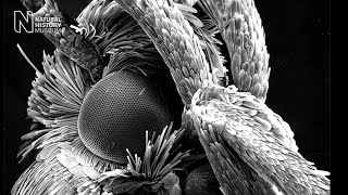
Scientists have imaged the brains of bumblebees in unprecedented detail, revealing the regions linked to learning and memory.
Museum researchers and collaborators used the Museum's micro-CT facility to scan the insects' brains in 3D, allowing them to see minute structures that would be damaged if the brains were removed.
These structures, in particular the 'mushroom bodies', are thought to help bees remember where to forage for pollen. Given the important role bees play in pollinating crops, understanding how their brains work - and how they may malfunction - is crucial.
'It's a fantastic way to look inside insect brains,' says Dr Richard Gill, from Imperial College London, who collaborated on the research. 'We can look at the brain as it naturally sits in the bee's head, without the human error of having to extract it.'
Micro-CT scanning uses X-rays to produce thousands of images of an object, which can then be assembled into a 3D model. It works in the same way as CT scanning, which is often done in hospitals, but at a much higher resolution - a bumblebee's brain is just 0.0002% the size of a human brain.
By fine-tuning this process, the research team managed to increase the resolution even further, allowing them to see details just five micrometres across - half the size of a human red blood cell.
This level of detail is important, as the team hopes to investigate the connection between behaviour and brain changes in lab-trained bumblebees. As the bees learn and remember, researchers believe that their brain structures change in size.
'With older techniques, the sizes of these structures could not be accurately measured and compared between bees,' says Dylan Smith, the lead author of the paper and a PhD student at the Museum and Imperial. 'The structures are so small that tiny errors in measurement can lead to wrong conclusions.
'This new technique allows structures to be isolated, examined, and measured in greater detail than ever before.'
A 3D reconstruction of a bumblebee brain that was scanned by the research team
The team believes the technique could now be used to investigate the effect of stressors on bee brain development and behaviour. Stressors such as disease, trauma and agricultural chemicals have been suggested as causes of a worldwide decline in bee populations.
This research will require a global effort, and by publishing their work, the team hopes that other teams around the world will be able to adopt their technique.
'More and more labs around the world have CT scanning equipment,' says Dr Farah Ahmed, who heads the Museum's CT facilities and who also collaborated on the research. 'But it's also a case of having the right techniques at your disposal.
'We hope that by sharing our process, we can help other labs in pushing this vital research forwards.'
The research was published in Nature's Scientific Reports.

Experts argue that changing the way we use land could help bees even more.

Discover the flowers that go to extraordinary lengths to attract insect pollinators.
Find out how the Museum’s traditional artworks and images made using modern techniques help shape our understanding of the natural world.

Give solitary bees a home by making this simple, DIY bee hotel.