Skip navigation

The NaturePlus Forums will be offline from mid August 2018. The content has been saved and it will always be possible to see and refer to archived posts, but not to post new items. This decision has been made in light of technical problems with the forum, which cannot be fixed or upgraded.

We'd like to take this opportunity to thank everyone who has contributed to the very great success of the forums and to the community spirit there. We plan to create new community features and services in the future so please watch this space for developments in this area. In the meantime if you have any questions then please email:

Fossil enquiries: esid@nhm.ac.uk
Life Sciences & Mineralogy enquiries: bug@nhm.ac.uk
Commercial enquiries: ias1@nhm.ac.uk

1

Jambo from Mwanza, Tanzania


It's my last week here and as much as I have enjoyed the fieldwork I can't wait to get back home. But there are a few more days of work to do first!


Back to blood-fluke fieldwork


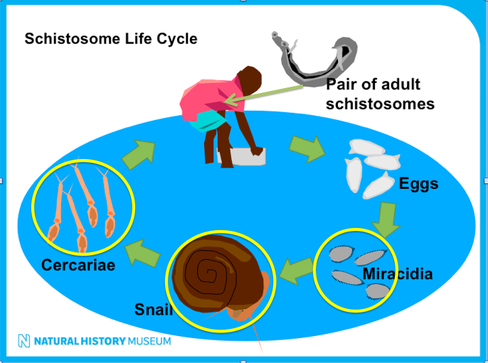
This time I want to tell you about our snail collecting work. Snail in Swahili is called Konokono. The snails we are interested in are aquatic, pulmonate little dudes belonging to the Biomphalaria genus.


They are the intermediate host of Schistosome mansoni, the blood fluke species responsible for intestinal schistosomiasis and it's detrimental health consequences in humans (see previous post - the Blood Fluke story).


We collect these snails in order to study the blood fluke parasites they carry.


The collecting process involves:


  • Scooping for snails on banks of Lake Victoria. We use protective waders to prevent blood fluke infection from the water.
  • Carrying the snails back to the lab, where we use microscopes to identify schistosome parasites.
  • Documenting the infected snails, which will be taken back to the Museum for DNA analysis.


aquatic snail Biomphalaria species, part of schistosome life cycle.jpg

Aquatic snail of the Biomphalaria genus, host to the human blood fluke Schistosoma mansoni, the causative agent of schistosomiasis.


collecting Muriel&team.jpg

Muriel and the team scooping for snails on the banks of Lake Victoria.


Revo&James.jpg

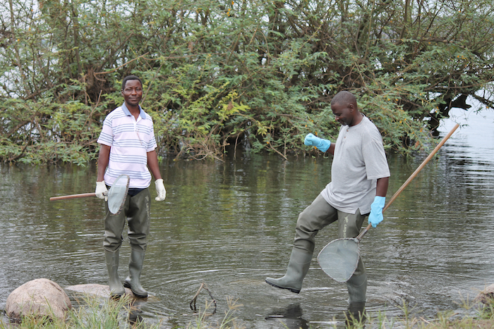
Mr Revocatus and Mr James with the snail scoops and protective clothing (hip waders) to prevent blood fluke infection from the water. Credit Fion Allan.

kids by fishing boats C Fiona.jpg

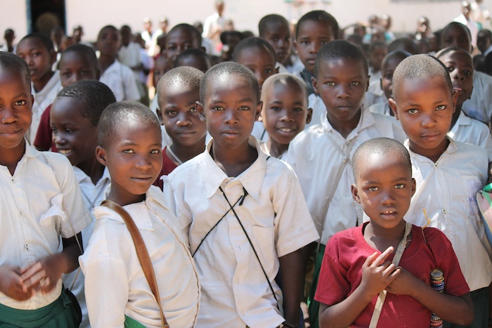
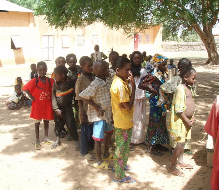
Village kids from local fishing village. Credit Fiona Allan.


Revo carring dregde Magu.jpg

Mr Revocatus carrying the dredge to our next snail site. Yes this is a beach on Lake Victoria. Not the sea!


James&dredge.jpg

Mr James with the dredge getting ready to collect those lake bottom snails. Credit Fiona Allan.


snails in dredge Cd Fiona.jpg

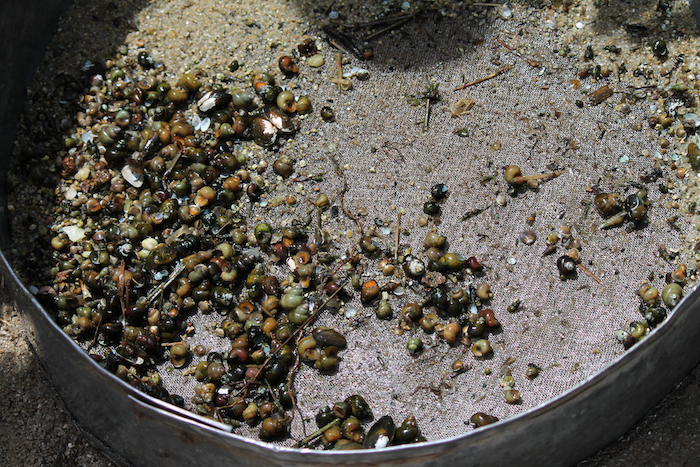
Dredged up snails from the lake bottom. Now we have to find the small Biomphalaria species we are after. Credit Fiona Allan.


avoiding the cow.jpg

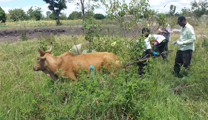
Sometimes we have to work around the local fauna.


Ducks are agathering cd Fiona.jpg

More local fauna. Credit Fiona Allan.


drying fish.jpg

Activities by the snail collecting sites. This lady is drying small fish in the sun.


Untitled-2.jpg

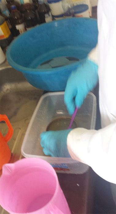
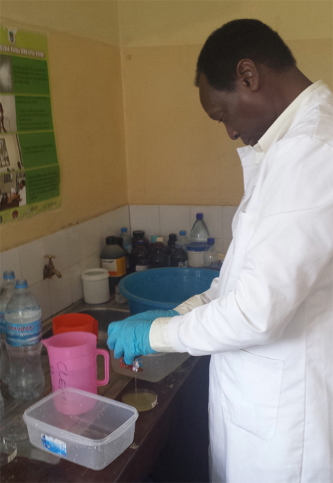
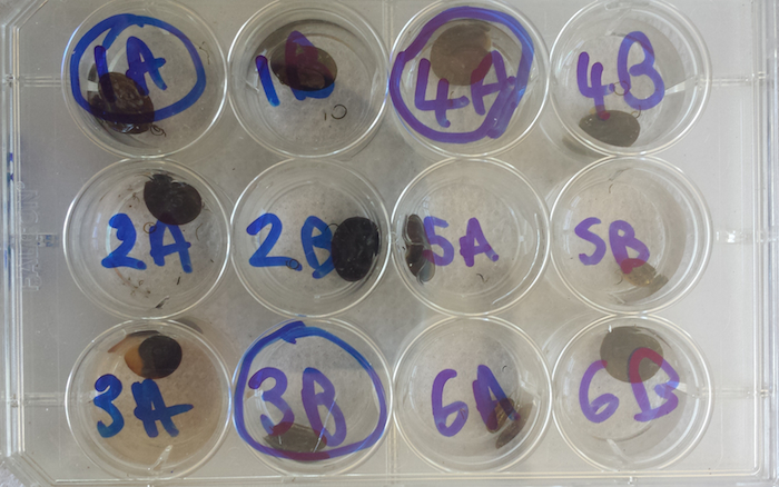
Back in the lab, we sort through all our collected snails, put them in water and check for schistosome parasites.


individual snails.jpg

Biomphalaria snails in individual wells with water. We check each well for the presence of the parasite larval stage, cercariae.


Schistosoma mansoni cercaraie&Biomphalaria sudanica.jpg

Blood fluke larvae (cercariae) under the microscope - those little white things in the water. They're looking for humans to infect!


identifying infected snails.jpg

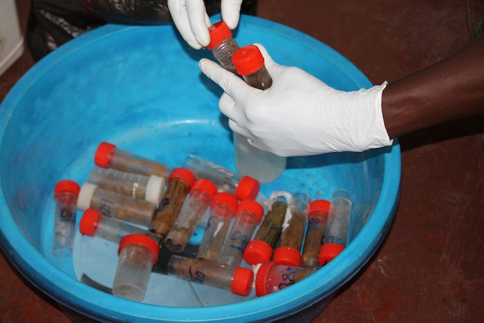
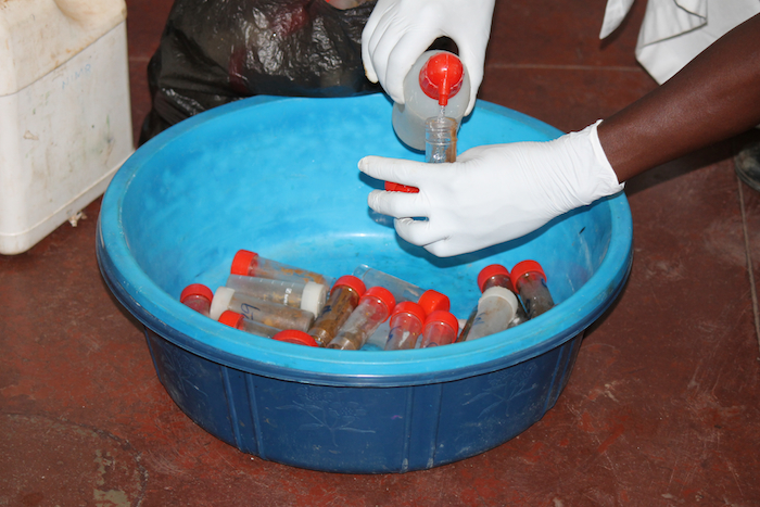
Identifying infected snails and giving them an ID number. We then preserve the snail in ethanol and bring them back to the Museum for genetic barcoding (species identification).


End of a hard week -Muriel&James.jpg

After a hard days work, Muriel and James getting ready to tuck into some food.


Untitled-1.jpg

Me about to eat some Wali na Samaki (rice and fish).

0

Jambo from Tanzania,


I realise I'm a bit late with this post so lets get straight to it. Just a warning though, this will be a rather disgusting post so get ready to be grossed out.


You'll remember from my previous post about our school visits that we collect stool samples from infected children. This is because we collect the miracidia larval stage that hatches out of the parasite eggs. And these eggs come out with stool.


The blood fluke life cycle - a recap


Schistosoma. The worm pair releases schistosome eggs into the blood system. The eggs pierce through the wall of the intestinal/urinary tract and exit the host when he/she defecates or urinates. They reach fresh water and hatch out into a larval stage called miracidia.


Life Cycle Screen shot 2014-04-25 at 16.35.40.jpg

Life cycle of Schistosome, blood fluke parasite and the specimens we collect during our fieldwork (circled in yellow).


So in order to collect miracidia we need stool from infected children. Diagnosis of infection is achieved using the Kato Katz method: a specimen of stool viewed on a microscope slide. If schistosome (blood fluke) eggs are observed in the stool specimen then the person is infected with at least one pair of schistosomes. For more information on diagnosis have a look at this video.


Collecting eggs from stool


Once we know which kids are infected we go to the schools and get stool samples (see previous post). We take these back to the lab and then a long process of stool filtering begins. We filter the stool for schistosome eggs, these we place in water and light. This induces them to hatch out into miracidia. We collect the miracidia onto special cards that store their DNA. We transport these back to the UK.


We use a pair of filters called Pitchford funnels (devised by Pitchford & Visser). The inner smaller funnel has bigger pores that allow the schistosome eggs to pass through but stops larger pieces of stool. The outer funnel is made of a finer mesh with pores that stop schistosome eggs from going through, this allows us to pour lots of water through the funnel thereby washing the eggs of stool material that may stop them from hatching.


Untitled-1.jpg

Pitchford Funnels (devised by Pitchford & Visser). Credit Fiona Allan.


stool.jpg

Stool Samples. Credit Fiona Allan.


Untitled-2.jpg

Sieve to break up stool. Credit Fiona Allan.


Untitled-3.jpg

Using the sieve to break up the stool sample.


Untitled-4.jpg

Pouring stooly water through Pitchford Funnel. Credit Fiona Allan.


John-Anouk processing stool samples.jpg

Myself and Mr John processing stool samples. Credit Fiona Allan.


stool-formalin.jpg

Adding formalin to left over stool samples to kill of anything inside. These are disposed of safely later. Credit Fiona Allan.


Revocatus-stool.jpg

>Revocatus adding formalin to stool. Credt Fiona Allan.


Untitled-5.jpg

My Nagai releasing the eggs and some water into a petri dish.


petridishes.jpg

Petri dishes of eggs and water. Waiting to hatch.


Untitled-6.jpg

Fiona starts checking for miracidia swimming in the petri dish.


NIMR lab-james-fiona.jpg

Fiona and James in the lab in National Institute for Medical Research in Mwanza.


Sometimes out in rural areas where we use local hospitals to process our samples things can go wrong, such as a power cut. No electricity means no light through the microscope. Thankfully we rise to the challenge and strap our head torches round our microscopes as an alternate source of light. Not quite as clear but it still works.


Untitled-7.jpg

Even a power cut will not stop us, we use our head torches as a light source and continue working.


So that's it for now. Tune in for the next post - snail collecting on the banks of Lake Victoria.

0

Jambo (hi) from Tanzania!


We are now into our second week of the trip and the blood fluke parasite collection is going well. A few logistical hiccups but nothing we can’t handle (so far).


Last week we went to a few schools to collect schistosomes from infected children. Just to recap why and what we are collecting from schools, here is the life cycle with the stages we are collecting on this trip:


Life Cycle Screen shot 2014-04-25 at 16.35.40.jpg

The life cycle of blood flukes, Schistosoma, involving a vertebrate (e.g. human) host and an aquatic snail host. Transmission is through contact of infested freshwater. The yellow circles are the stages and specimens we collect when doing fieldwork.


So we have the delightful job of collecting the larval stage, called miracidia, that hatch out from eggs. How do we do this? We go into a school, collect stool samples from infected children and filter out the eggs. We then put them in some water in sunlight and wait for them to hatch. I will explain this in more detail in a subsequent post on lab work. For now let's stick to the first stage: visiting schools.


We visit state primary schools in the Mwanza region of Lake Victoria. To get to these schools we sometimes have to drive for hours through dirt tracks. All sorts of obstacles occur but the most common one is this: cattle!


cattle.jpg

On our way to a school, a herd of cattle, goats and sheep block our path.


When we arrive we visit the head teacher and get a proper greeting from the school. The teacher then calls out our selected students - the ones we know are infected from a previous survey, more on this later.


school.jpg

Children were practicing singing, dancing and music on the day we arrived. Credit: Fiona Allan.


kids.jpg

Up close and personal, the kids stare at us. Eventually we do get them to smile. Credit: Fiona Allan.


We’re a small team: two scientists from the Museum (Fiona and myself) and 3 research technicians from the National Institute for Medical Research in Mwanza - Mr John, Mr Nagai and Mr James. As well as our trusted driver – Mr Lenard.


team.jpg

The team, Mr John, Mr Nagai and me. Getting our gloves on and our kit ready. Credit: Fiona Allan.


fiona-allan-resize.jpg

My colleague Fiona Allan, a brilliant schistosome expert and our trip’s photographer.


anouk-resize.jpg

Me holding a football I am about to present to the headteacher as a present. Credit: Fiona Allan.


James with the kids .jpg

Mr James is teaching the children how to give us a stool samples and most importantly to wash their hands afterwards! Good hygiene practice!


We give the kids a container to put a stool sample, and some toilet paper. They run off to the latrines and come back with a full container. How they are able to poop on demand always amazes me. We label the containers with unique identification numbers for each child. And then go back in the lab to process the samples. All the children in the school receive treatment a couple of weeks later. We always treat any infected child!


stool collecting.jpg

Mr Nagai and Mr John handing out toilet paper to the kids. Credit: Fiona Allan.


sample containers.jpg

The children all grab for a container for their stool sample.


latrines.jpg

School latrines. Credit: Fiona Allan.


washing hands.jpg

Mr James supervises the hand washing.


kichocho.jpg

We were very happy to see this in some of the schools: a warning about schistosomiasis, called Kichocho in Kiswahili, and an explanation about the life cycle. Credit: Fiona Allan.


abacus-rezize.jpg

Some shots from the school. A little girl with a necklace of bottle tops, this actually serves as a abacus in the schools. Credit: Fiona Allan.


Abacus.jpg

Local abacus, device for learning arithmetic. Credit: Fiona Allan.


play.jpg

Kids playing in front of a typical Mwanza rock.


This time I came with some gifts for the schools. Back in the UK I decided to get a football for each school. The footballs they use are often just rags and plastic wrapped into a tight ball and tied together, or completely deflated punctured balls. So I went shopping at Altimus. The staff and manager were curious about why I wanted 16 footballs. When I explained they very kindly gave me a generous discount. So this is a thank you to Altimus!


football.jpg

Kids playing football with their old cloth ball.


footballs.jpg

The new football next to the old football. You can see why the teachers and kids are delighted with the gift. Thank you Altimus.


netball-resize.jpg

Girls playing basketball with the new ball from Altimus.


school TZ .jpg

Time to say Asante (thank you) and Kwaheri (good bye).


That's it for today. Next post - what do we do with poo and how to go parasite fishing with a microscope.


Asante sana (thank you very much in Swahili).

0

The blood fluke story

Posted by Anouk Gouvras May 1, 2014

Hello super-fly and parasite enthusiasts. Time for blog post 2, which is coming to you from the Mwanza region of Tanzania, bordering the banks of Lake Victoria. My colleague and I are here to collect specimens of the blood fluke Schistosoma from infected humans and snails.


Infection with the blood fluke Schistosoma causes a disease called Schistosomiasis (aka Bilharzia). Over 200 million people worldwide are infected with over 700 million people living at risk of infection. Over 80% of infected people live in sub-Saharan Africa. It is a disease of low socio-economic status, affecting the poorest communities and most neglected, vulnerable people. Infants and children are especially prone to infection due to their less developed immune system.


school children in West Africa 700p.jpg

Children in a school in Niger, West Africa, queuing to be tested for schistosomiasis. The little boy at the front is showing the swollen liver and spleen symptom. A result of being infected with schistosomiasis.


There are two forms of the disease, depending on the species of the infecting schistosome worm:


Intestinal Schistosomiasis

  • diarrhoea, bloody stool
  • anaemia, stunted growth
  • enlarged liver and spleen
  • severe damage to the liver leading to liver fibrosis


Urogenital schistosomiasis

  • blood in urine, painful urination
  • anaemia, stunted growth
  • damage to the genitals, kidneys and bladder
  • bladder cancer
  • increased risk of sexually transmitted diseases like HIV


bloody urines.jpg

Urine samples from children infected with Schistosoma haematobium, the urogenital form of schistosomiasis. The red colour indicates blood seeping out with the urine due to the damage done by the schistosome eggs.


To help fight schistosomiasis we need to understand the complex life cycle of Schistosoma, which involves a vertebrate (in this case human) host, a snail host and transmission via water contact.


The blood fluke life cycle


Lets start with a worm pair living inside a little boy in sub-Saharan Africa. The worm pair resides in the blood system of the little boy, either around the intestinal tract or around the urinary tract depending on the species of Schistosoma.


The worm pair releases schistosome eggs into the blood system. The eggs pierce through the wall of the intestinal/urinary tract and exit the boy when he defecates or urinates. They reach fresh water and hatch out into a larval stage called miracidia. These infect a specific aquatic snail species and reproduce asexually, creating thousands of clonal larval stages called cercaraie.


Cercaraie leave the snail to locate and infect a human by piercing through exposed skin in the water. They travel to the liver via the blood system and there they mature into adult worms, ready to reproduce and continue the life cycle (see diagram below).


Life Cycle Screen shot 2014-04-25 at 16.35.40.jpg

The life cycle of blood flukes, Schistosoma, involving a vertebrate (e.g. human) host and an aquatic snail host. Transmission is through contact with infested freshwater. The yellow circles are the stages and specimens we collect when doing fieldwork. Credit: Aidan Emery.


Schistosoma blood fluke worm pair700p.jpgA schistosome worm pair. The fat male carries the thinner female worm folded in a little groove where he feeds and shelters her whilst she produces eggs. The worm pair lives inside the veins of animals.


The schistosome species I work on (Schistosoma mansoni) causes intestinal schistosomiasis. It lives in the vein blood system of the liver and intestinal tract of humans. The adult worms themselves don’t cause much harm but it is the eggs they produce that cause the disease, by:


  • Piercing the barrier between the blood system and the intestinal wall = bloody diarrhoea and painful cramps.
  • The eggs that don’t make it out get trapped in organ tissues, causing the immune system to overreact and damage the surrounding human tissues.
  • More worm pairs = more eggs = more damage to the organs and the host. This is what causes the chronic and more severe aspects of the disease such as kidney failure, bladder cancer and liver fibrosis in adulthood.


Thankfully there is an effective oral drug called Praziquantel that kills the adult worms. However it cannot prevent children from becoming infected. So in areas where there is no clean piped water or a sewage system, the local water bodies such as Lake Victoria are the only sources of water for the local population. People have no choice but to fish, wash, bathe and collect water from these schistosome-infested waters and therefore are reinfected quickly.


Treatment needs to be repeated regularly to avoid heavy worm numbers and high egg outputs. Regular treatment means controlling the disease but does not mean eliminating it. For this we need more research to develop better tools to fight the disease. There is also a major the worry that the parasite will become resistant to the drug. If the parasite develops resistance and the drug stops working there is currently no alternative treatment.


West Africa schistosome transmission site and local water collection point.jpg

Clean water is not available so local water-bodies are used, such as this irrigation canal in Niger, West Africa.


We are researching the impact of human treatments on the parasite population. This will reveal how the parasite is responding to ongoing treatment programmes, if the drug is working effectively and if there are any warning signs regarding drug resistance.


That’s it for now. Coming up, a visit to the schools to collect stool samples from infected children. Disgusting work but someone’s got to do it.