Skip navigation

The NaturePlus Forums will be offline from mid August 2018. The content has been saved and it will always be possible to see and refer to archived posts, but not to post new items. This decision has been made in light of technical problems with the forum, which cannot be fixed or upgraded.

We'd like to take this opportunity to thank everyone who has contributed to the very great success of the forums and to the community spirit there. We plan to create new community features and services in the future so please watch this space for developments in this area. In the meantime if you have any questions then please email:

Fossil enquiries: esid@nhm.ac.uk
Life Sciences & Mineralogy enquiries: bug@nhm.ac.uk
Commercial enquiries: ias1@nhm.ac.uk

Super-flies and parasites

8 Posts tagged with the entomology tag
0

On 25 June the Museum will open its doors to a special event in celebration of the international and global commitment between countries, industry, charities and academia to work together against Neglected Tropical Diseases (NTDs). This commitment was first agreed upon in London in 2012 and has since been termed the London Declaration On NTDs.


By joining forces to fight NTDs the world would achieve a huge reduction in health inequality paving the way to sustainable improvements in health and development especially amongst the worlds poor. The 25 June sees the launch of the third progress report, 'Country Leadership and Collaboration on Neglected Tropical Diseases'. A pragmatic overview of what has been done, what has worked, what hasn't and what key areas still need to be achieved.


The Museum is thrilled to be participating in this event, having a long-standing history in parasitic and neglected tropical disease research. As both a museum and an institute of research our mission is to answer questions of broad significance to science and society using our unique expertise and collections and to share and communicate our findings to inspire and inform the public. We are excited to be hosting a day of free public events on Neglected Tropical Diseases.


What are NTDs?

Neglected Tropical Diseases are termed in this way because they infect hundreds of thousands to millions of people, predominantly the world's poorest and most vulnerable communities, and yet receive comparatively little funding for basic, clinical or drug-development research and even less attention from governments, people and the media of affluent countries. Until now!


In total the WHO has identified 17 diseases or groups of diseases that fall within this category.


NTD slides from Bonnie Webster.jpg

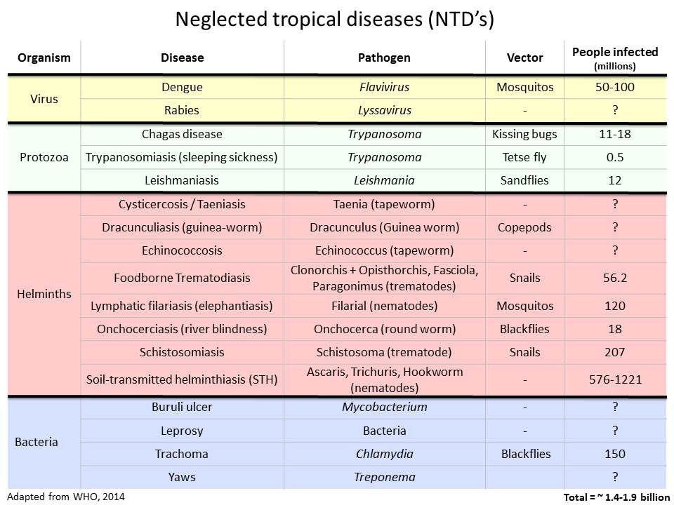
World Health Organization has identified 17 Neglected Tropical Diseases. 10 of these have been targeted for control and elimination by 2020


The 10 selected by the WHO for control and elimination by 2020 are:


  1. Onchocerciasis (aka river blindness): A blood worm infection transmitted by the bite of infected blackflies causing severe itching and eye lesions as the adult worm produces larvae and leading to visual impairment and permanent blindness.
  2. Dracunculiasis (aka Guinea-worm disease): A roundworm infection transmitted exclusively by drinking-water contaminated with parasite-infected water fleas. The infection leads to meter-long female worms emerging from painful blisters on feet and legs to deposit her young. This leads to fever, nausea and vomiting as well as debilitating secondary bacterial infections in the blisters.
  3. Lymphatic filariasis: A blood & lymph worm infection transmitted by mosquitoes causing abnormal enlargement of limbs and genitals (elephantiasis) from adult worms inhabiting and reproducing in the lymphatic system.
  4. Blinding trachoma: A chlamydial infection transmitted through direct contact with infectious eye or nasal discharge, or through indirect contact (e.g. via flies) with unsafe living conditions and hygiene practices, which if left untreated causes irreversible corneal opacities and blindness. Trachoma is the leading cause of blindness in the word.
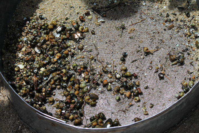
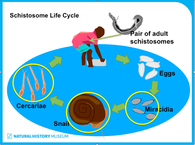
  5. Schistosomiasis (aka bilharzia): A blood fluke infection transmitted when larval forms released by freshwater snails penetrate human skin during contact with infested water. The infection leads to anaemia, chronic fatigue and painful urination/defaecation during childhood, later developing into severe organ problems such as liver and spleen damages, bladder cancer, genital lesions and infertility.
  6. Visceral leishmaniasis (aka Kala azar): A protozoan blood parasite transmitted through the bites of infected female sandflies which attacks internal organs which can be fatal within 2 years. 
  7. Soil-transmitted helminths: A group on intestinal worm infections transmitted through soil contaminated by human faeces causing anaemia, vitamin A deficiency, stunted growth, malnutrition, intestinal obstruction and impaired development.
  8. Leprosy: A complex disease caused by infection mainly of the skin, peripheral nerves, mucosa of the upper respiratory tract and eyes.
  9. Chagas disease: A life-threatening illness caused by a blood protozoan parasite, transmitted to humans through contact with vector insects (triatomine bugs), ingestion of contaminated food, infected blood transfusions, congenital transmission, organ transplantation or laboratory accidents.
  10. Human African trypanosomiasis (aka sleeping sickness): A protozoan blood parasitic infection spread by the bites of tsetse flies that is almost 100% fatal without prompt diagnosis and treatment to prevent the parasites invading the central nervous system.


They were selected because the tools to achieve control are already available to us and, for some, elimination should be achievable.


Take the Guinea Worm:


Guinea worm Peter Mayer.jpgGW (SKnopp).jpg

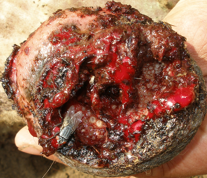
Guinea worm infection - from over 3.5 million people infected in the 80s to less than 130 cases in 2014. Set to be second human disease to be eradicated after smallpox (photo credits David Hamm&Peter Mayer)


In the 1980s over 3.5 million people were infected with Dracunculiasis (i.e. Guinea worm disease), with 21 countries being endemic for the disease. Now, thanks to the global health community efforts and extraordinary support from the Carter Center, only 126 cases were reported in 2014 and only 4 endemic countries remain: Chad, Ethiopia, Mali and South Sudan! If the WHO goal of global eradication of Guinea Worm by 2020 is met then Dracunculiasis is set to become the second human disease in history to be eradicated (the first, and only one, being smallpox). Not bad for an NTD! But there are still challenges!


At the Museum we have a long history of working on health related topics. Indeed our founding father Sir Hans Sloane was a physician who collected and identified plants from all over the world for the purpose of finding health benefits - in fact he developed chocolate milk as a health product.


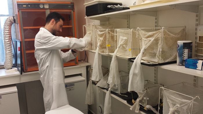
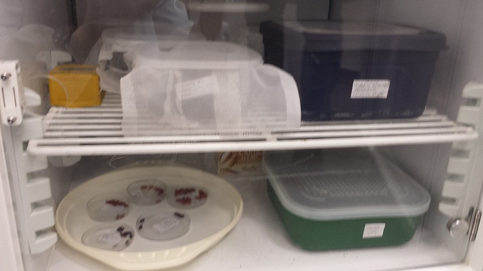
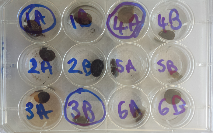
Today we have a vast and biologically diverse collection of parasites and the insects/crustaceans/snails/arachnids that carry and transmit them. These are used by researchers both in the museum (such as myself and colleagues) but also internationally through collaborative work.


ZEST Zanzibar.jpg

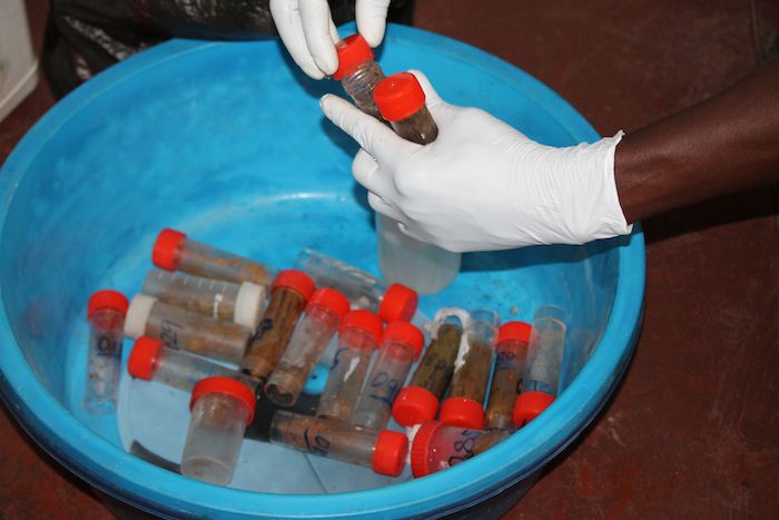
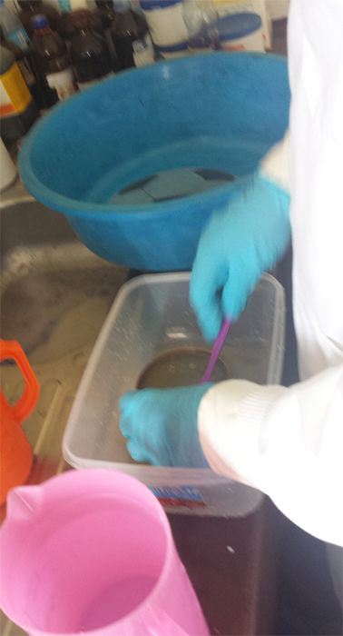
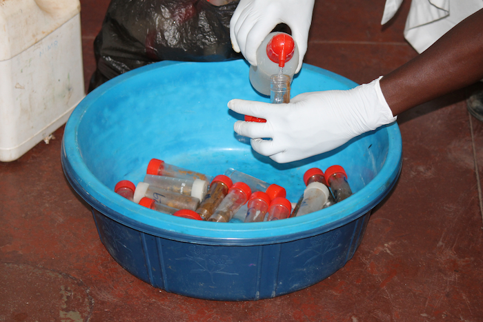
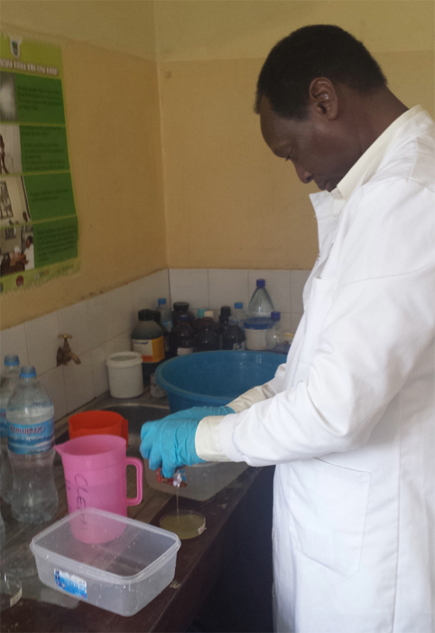
Collaboration is key - Zanzibar Elimination of Schistosomiasis Transmission (ZEST) programme key players: the Zanzibar Ministry of Health, Public Health Laboratories Pemba, the World Health Organization, SCI, SCORE, Swiss TPH, NHM and others


We are immensely proud of our collections and the work we do in this field especially of the biological information we can contribute to health programmes in endemic countries. One of our most exciting contributions is to the Zanzibar Elimination of Schistosomiasis Transmission (ZEST) programme where we are working in collaboration with the Zanzibar Ministry of Health, various NGOs, the World Health Organization and the local communities to identify and implement the best tools and methods to achieve schistosomiasis elimination in Zanzibar. This would be the first time a sub-Saharan African country would achieve schistosomiasis elimination. Fingers-crossed we are up to the challenge! You can read more about this project in an earlier post on our Super-flies and parasites blog


On Thursday we are bringing out our Parasites and Vectors specimens to showcase them to the public galleries and answer any questions relating to these fascinating yet dangerous organisms. Our wonderful scientists and curators will be on hand to talk to people about our collections and research as will collaborating scientists from the London Centre of Neglected Tropical Disease Research who will talk to you about the diseases and the challenges faced to achieve the WHO 2020 goals. Please do pop by and say hello, come and look at our specimens and help us raise awareness of these devastating diseases and the fight to control and eliminate them.


Fieldwork_TZ_FionaAllan_SCAN.jpg

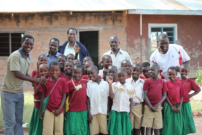
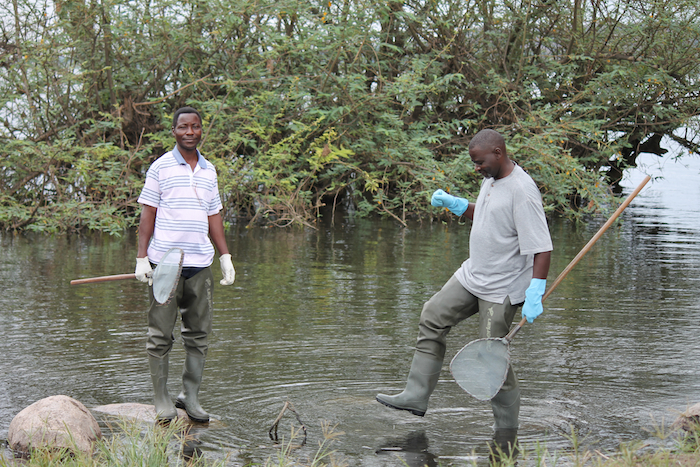
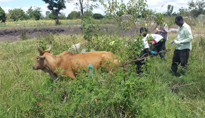
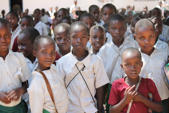
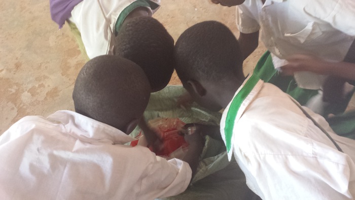
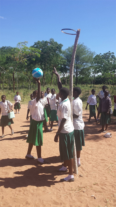
We are working together with schools, communities, government and research institutes to fight Neglected Tropical Diseases. Schistosomiasis fieldwork photo with the team from the National Institute for Medical Research in Tanzania

0

Hello Super-flies and Parasites fans!


We are back with all things nasty from the Parasites and Vectors division here at the Museum. There have been some exciting developments in the New Year, most importantly the launch of the Museum’s brand new website!


This is another ‘Forever Flies’ series of blog posts, bringing you news from the Museum'sforensic entomologygroup.

green-bottle fly.jpg

Forever Flies is our forensic entomology blog series. This image shows a carrion-eating greenbottle blowfly.


Forensic Entomology

You will remember from my previous Forever Flies post that forensic entomology is the study of the insects and arthropods found at a crime scene. The most common role for Museum forensic entomologists is establishing a minimum time since death in suspicious cases, by analysing the carrion insects on the body.


2014-10-16 Gross maggots with adult.jpg   issue2forensic3_large.jpg

Blowflies use the bodies of dead animals to grow and develop. The rate at which they do this, going from egg to larva to pupa to adult fly, is pretty consistent and depends largely on ambient temperature. Forensic entomologists use this to determine the minimum post-mortem interval (PMImin), which helps crime scene investigators determine approximate time-of-death.


Thanks to entomological expertise (Greek – entomo = insect, logos = knowledge) scientists can collect insects from a corpse and/or crime scene, determine what stage in their life cycle the insects have reached and, using their knowledge on the duration of each stage of the insects’ life cycle, determine how long ago the parent insect laid her eggs on the corpse.


This gives an incredibly useful estimate of the minimum amount of time this body has been dead (minimum post-mortem interval - PMImin), which helps crime scene investigators determine approximate time-of-death. The more accurate this minimum post-mortem interval is, the more accurate the time of death can be. Knowing time of death can focus the police investigation and suggest the likelihood of a suspect’s involvement.


Scientists can also use these insects to determine if the body has been moved since death and how long a body was exposed above ground before burial.


Metamorphosis in pupae


Flies spend over 50% of their developmental life in the pupae stage, protectively encased inside a hard shell (called a puparium) where they slowly transform from a maggot into a fly in a process called metamorphosis (Greek again - Meta = change, morphe = form).


A puparium looks quite bland and boring but underneath there are all sorts of wonderful things going on. Scientists can remove the shell and, using traditional microscopy, take a look at the fascinating changes of metamorphosis. But this process does destroy the pupa sample, making it difficult to work out how long it takes for the pupa to go through the different stages of metamorphosis.


Scientists know that the length of time metamorphosis takes to complete really depends on temperature, the question is can we use our knowledge of the process to pinpoint a more accurate estimate of PMImin?  What forensic scientists need is a standardised method to work out:

  1. At what stage in the metamorphosis process is the pupa
  2. how long did it take to reach this stage


If these two points can be determined then scientists can provide a far more accurate PMImin.


The ‘MORPHIC’ project


Dr Daniel Martin-Vega, a forensic entomologist, has joined the Museum from the University of Alcalá in Spain to research carrion fly pupae and to develop a standardised protocol for aging pupae (as in determining their age) that can be used by forensic scientists. This project is called MORPHIC and is funded by the European Commission through a Marie-Curie fellowship.


It sounds all neat, logical and tidy but there is A LOT of work and dedication involved!


For this projectDaniel is raising two species of the carrion-loving blowflies, the greenbottle blowfly Lucilia sericata and the bluebottle blowfly Calliphora vicina. The flies live in netting covered cages, where they feed and reproduce whilst he monitors them.


Daniel feeding flies_resized2.jpgDaniel showing me the Diptera (insect) culture room. Each netting-covered box has a species of carrion blowfly in it. He is researching the pupae of these flies to see if he can improve the estimate of  PMImin and thus improve the information given to crime scene investigators.


He also has to collect the post-feeding maggots and place them in a box with some nice clean soil for them to happily grow until they are ready to start the metamorphosis process. These boxes are then placed in a cabinet kept at a specific temperature. Since the rate of metamorphosis largely depends on temperature it is very important the Daniel can control this environmental factor in order to document the rate of change at different temperatures.


where do you keep the maggots_resized2.jpg

The maggot house! This is a comfy box with soil where maggots crawl around and prepare to pupate. When the maggots start pupating Daniel has to come in every 6 hours or so to monitor and collect them for his research


puppa&maggots_resized.jpg

Blowfly maggots and pupae.


Once the maggots start to pupate Daniel has to collect the pupae:


I come in every 6 hours when the maggots start to pupariate in order to collect blowfly pupae at 6-hour intervals during the first 48 hours after puparium formation (the period when the greatest morphological changes of metamorphosis occur). Luckily, I only do this from time to time. After that, the collection of pupae is just daily until the adult flies’ emergence.

picking out pupae_resized.jpg

Daniel sieving out the pupae from the box.


Maggots&pupae_resized.jpg

Maggots and pupae, oh my!


Watch those maggots wriggle about!


He then has to sieve out the pupae from the soil and carefully place them in a petridish labelled with the blowfly species name, the date collected and the time collected. These petridishes are also placed in the special temperature-control cabinet.


Fly puppa sorted_resized.jpg

Daniel has separated out the pupae of different species of blowfly. Each petridish with pupae has the species name, the date collected and the time collected.


where do you keep the maggots_resized.jpg

The petridishes are kept at a specific temperature. Since the rate of metamorphosis largely depends on temperature it is very important the Daniel can control this environmental factor.


Daniel uses the Museum’s wonderful micro-computed tomography (micro-CT) scanner to take detailed images of the inside of the pupae without destroying them. A micro-CT scanner is a type of X-ray scanner that produces 3D images, much like a hospital CAT scanner, but at a much smaller scale and a higher resolution. The results are like 3D microscope images! 


Thomas Simonsen and Daniel Martin-Vega analysing CT images of 5 pupae.jpg Thomas Simonsen and Daniel Martin-Vega operating Micro-CT scanner.jpg

Daniel with colleague Dr Thomas Simonsen using the Museum’s micro-CT scanner to look at 3D images of blow-fly pupae. The micro-CT scanner uses x-ray technology to produce 3D 'microscopy' images at high resolution without damaging the sample.


By using the Museum’s micro-CT scanner Daniel can take these detailed images at specific time points of the metamorphosis process.  He will then have a catalogue of images of the blow fly pupal development at specific temperatures. This catalogue of images will be used to develop a standardised tool to determine the age of blow fly pupae. Then when pupae are collected from a crime scene, they can be compared to this catalogue and scientists will be able to determine how long the fly has been in its pupal stage. Giving scientists a more accurate estimate of PMImin! Ta daaaaa!


C_vicina-48h.jpgC_vicina-216h.jpg

Micro-CT scanner images of a bluebottle blowfly Calliphora vicina pupa. The one on the left is at 48 hours, the one on the right at 216 hours. You can see the difference in development between the two pupa images.


Micro-CT scan of blowfly pupa.jpg

Dorsal micro-CT scanner image of a blowfly pupa.


I hope you enjoyed this post. If you fancy a stab at a bit of CSI work why not check out the Museum's Crime Scene Live After Hours events.

0

Hello Super-flies & Parasites fans!


This time we are departing from the familiar world of blood flukes and having a look at something new and exciting: Welcome to the ‘Forever Flies’series of blog posts. I’ve really enjoyed writing this bit as it’s totally new to me and I’m learning so much from our wonderful scientists in the Forensic Entomology group of the Parasites & Vectors division here at the Museum.


About forensic entomology


I think I mentioned forensic entomology way back in the first ever Super-flies and Parasites post. But to refresh our memories and delve a bit deeper here’s an explanation taken from the group’s website:

'Forensic entomology is the study of insects and other arthropods (ie spiders, mites) in a situation where a crime may have been committed. The insects recovered from a crime scene can provide vital information for the investigating team. The most common role for Museum forensic entomologists is establishing a minimum time since death in suspicious cases, by analysing the carrion insects on the body.'


Flies use the bodies of dead animals to grow and develop, fulfilling a vital nutrient recycling role in an ecosystem. They can turn one vertebrate body into thousands or millions of flies, which are then fed on by other animals - insects, frogs and birds for example. The rate at which they do this, going from eggs to larvae to pupa to adult fly, is pretty consistent.


Knowing enough about the species of flies we can exploit this information when they develop on corpses. By determining how long the insects have been feeding on the tissues of the corpse, we can determine the length of time elapsed since flies found the body; thereby providing crime scene investigators with a minimum post-mortem interval.


Some flies however don’t hang about and wait for death, they prefer feeding on live animal tissues, and can cause a horrible disease called myiasis, a major economic and animal welfare problem.


2014-10-16 Gross maggots with adult.jpg

Female Bluebottle blow fly, Calliphora vicina, and maggots feeding on a dead pig.


Dr Martin Hall and colleagues within the Museum and around the world work together on the taxonomy and biology of flies that develop as larvae on living or dead vertebrate animals. Their expertise and research greatly contributes to the control of a painful and damaging disease, myiasis, but also to the field of forensic entomology, helping CSIs determine crucial information from a crime scene.


So there you have it, some real life crime scene investigation stuff! These are the guys CSIs turn to for help! (Cue CSI theme song!)


The beauty of maggots lies in their mouthparts


I asked Martin for a little blurb to get the Forever Flies series rolling and he sent me some surprisingly beautiful photos of maggots! Here’s what he has to say about them:

To most people maggots are repulsive creatures; they all look much the same and have zero redeeming features.


2014-10-16 Gross maggots 1-1.jpg2014-10-16 Gross maggots 2.jpg

Maggots, thought of as repulsive creatures with zero redeeming features. Read on to see how they are transformed by confocal microscopy into beautiful works of art.


However, viewed under a microscope they become much more interesting, with a range of characters that can be used in discrimination, especially when you look at the business end of the maggot, its mouthparts!


It’s not so easy to ask a tiny 2mm long newly hatched maggot to 'open wide' to view the teeth, and traditionally we have been limited to viewing slide-mounted specimens by light microscopy. Scanning electron microscopy has its merits, but only for the external features. In normal light microscopy, imaging of these mouthpart structures is limited by problems of resolution, illumination and depth of field.


Lucilia sericata L1 - Light microscope high power.jpg

Greenbottle blowfly, Lucilia sericata light microscope high power image. With normal light microscopy the relationship of the sclerites of the cephaloskeleton (mouthparts) to each other is unclear.


At the Museum we have been using a laser confocal microscope for the first time to look inside these maggots to view the mouthparts, the so-called cephaloskeleton, in three dimensions. The mouthparts are crucial to the maggots in establishing themselves on their food source, be it a live animal or a decomposing corpse. The images produced by the confocal microscope rely on the autofluorescence of structures of the cephaloskeleton.


Lucilia sericata LI - Confocal microscope low power.jpg

Greenbottle blowfly, Lucilia sericata confocal microscope low power image - relationships are still unclear but, with the autofluorescence under laser light, the structures look so much more beautiful!


We were especially interested in the relationships of the small sclerites to each other and the so called 'hump', present in newly hatched larvae of Lucilia greenbottle blowflies but absent in Calliphora bluebottles.


Lucilia sericata L1 - Confocal microscope high powerWITH ARROW.jpg

Greenbottle blowfly, Lucilia sericata confocal microscope high power image: The structure relationships are becoming clearer (the 'hump' is arrowed) and this is finalised in the next image.


The 177 optical sections scanned by the confocal microscope enabled us to rotate and view this structure in three dimensions (see green false-coloured sclerite in image below) and see clearly for the first time how it relates to other structures.


In addition to their academic and practical value in identification, the images are also things of beauty in their own right and would not look out of place in an art gallery!


Lucilia sericata - Confocal high power - false colour sclerites named.jpg

Greenbottle blowfly, Lucilia sericata confocal microscope high power image and false colour sclerites. We have rotated the original to give three dimensionality. The red wavelength was selected, as this is the wavelength that gave most autofluorescence of the cephaloskeleton, to enable us to discard other structures and here false-colour has been added to show the different structures, including the 'hump' (in green) or epistomal sclerite.


The work was done at the Museum and involved Drs Andrzej Grzywacz (a visitor under the EC-funded SYNTHESIS project) and Krzysztof Szpila from the Nicolaus Copernicus University in Torun, Poland, collaborating with Tomasz Góral and Martin Hall. We used the Museum’s Nikon A1-Si Confocal Microscope.


For more information see the article published online in Parasitology Research on 19 September 2014.


By Dr Martin Hall


I hope you enjoyed the first 'Forever Flies' post. For more on flies head over to Erica McAlister's Diptera blog.


There will be more coming soon but just to let you know there may be a couple of weeks break whilst some important maintenance work is done to the site and my life


See you soon

1

Jambo from Mwanza, Tanzania


It's my last week here and as much as I have enjoyed the fieldwork I can't wait to get back home. But there are a few more days of work to do first!


Back to blood-fluke fieldwork


This time I want to tell you about our snail collecting work. Snail in Swahili is called Konokono. The snails we are interested in are aquatic, pulmonate little dudes belonging to the Biomphalaria genus.


They are the intermediate host of Schistosome mansoni, the blood fluke species responsible for intestinal schistosomiasis and it's detrimental health consequences in humans (see previous post - the Blood Fluke story).


We collect these snails in order to study the blood fluke parasites they carry.


The collecting process involves:


  • Scooping for snails on banks of Lake Victoria. We use protective waders to prevent blood fluke infection from the water.
  • Carrying the snails back to the lab, where we use microscopes to identify schistosome parasites.
  • Documenting the infected snails, which will be taken back to the Museum for DNA analysis.


aquatic snail Biomphalaria species, part of schistosome life cycle.jpg

Aquatic snail of the Biomphalaria genus, host to the human blood fluke Schistosoma mansoni, the causative agent of schistosomiasis.


collecting Muriel&team.jpg

Muriel and the team scooping for snails on the banks of Lake Victoria.


Revo&James.jpg

Mr Revocatus and Mr James with the snail scoops and protective clothing (hip waders) to prevent blood fluke infection from the water. Credit Fion Allan.

kids by fishing boats C Fiona.jpg

Village kids from local fishing village. Credit Fiona Allan.


Revo carring dregde Magu.jpg

Mr Revocatus carrying the dredge to our next snail site. Yes this is a beach on Lake Victoria. Not the sea!


James&dredge.jpg

Mr James with the dredge getting ready to collect those lake bottom snails. Credit Fiona Allan.


snails in dredge Cd Fiona.jpg

Dredged up snails from the lake bottom. Now we have to find the small Biomphalaria species we are after. Credit Fiona Allan.


avoiding the cow.jpg

Sometimes we have to work around the local fauna.


Ducks are agathering cd Fiona.jpg

More local fauna. Credit Fiona Allan.


drying fish.jpg

Activities by the snail collecting sites. This lady is drying small fish in the sun.


Untitled-2.jpg

Back in the lab, we sort through all our collected snails, put them in water and check for schistosome parasites.


individual snails.jpg

Biomphalaria snails in individual wells with water. We check each well for the presence of the parasite larval stage, cercariae.


Schistosoma mansoni cercaraie&Biomphalaria sudanica.jpg

Blood fluke larvae (cercariae) under the microscope - those little white things in the water. They're looking for humans to infect!


identifying infected snails.jpg

Identifying infected snails and giving them an ID number. We then preserve the snail in ethanol and bring them back to the Museum for genetic barcoding (species identification).


End of a hard week -Muriel&James.jpg

After a hard days work, Muriel and James getting ready to tuck into some food.


Untitled-1.jpg

Me about to eat some Wali na Samaki (rice and fish).

0

Jambo from Tanzania,


I realise I'm a bit late with this post so lets get straight to it. Just a warning though, this will be a rather disgusting post so get ready to be grossed out.


You'll remember from my previous post about our school visits that we collect stool samples from infected children. This is because we collect the miracidia larval stage that hatches out of the parasite eggs. And these eggs come out with stool.


The blood fluke life cycle - a recap


Schistosoma. The worm pair releases schistosome eggs into the blood system. The eggs pierce through the wall of the intestinal/urinary tract and exit the host when he/she defecates or urinates. They reach fresh water and hatch out into a larval stage called miracidia.


Life Cycle Screen shot 2014-04-25 at 16.35.40.jpg

Life cycle of Schistosome, blood fluke parasite and the specimens we collect during our fieldwork (circled in yellow).


So in order to collect miracidia we need stool from infected children. Diagnosis of infection is achieved using the Kato Katz method: a specimen of stool viewed on a microscope slide. If schistosome (blood fluke) eggs are observed in the stool specimen then the person is infected with at least one pair of schistosomes. For more information on diagnosis have a look at this video.


Collecting eggs from stool


Once we know which kids are infected we go to the schools and get stool samples (see previous post). We take these back to the lab and then a long process of stool filtering begins. We filter the stool for schistosome eggs, these we place in water and light. This induces them to hatch out into miracidia. We collect the miracidia onto special cards that store their DNA. We transport these back to the UK.


We use a pair of filters called Pitchford funnels (devised by Pitchford & Visser). The inner smaller funnel has bigger pores that allow the schistosome eggs to pass through but stops larger pieces of stool. The outer funnel is made of a finer mesh with pores that stop schistosome eggs from going through, this allows us to pour lots of water through the funnel thereby washing the eggs of stool material that may stop them from hatching.


Untitled-1.jpg

Pitchford Funnels (devised by Pitchford & Visser). Credit Fiona Allan.


stool.jpg

Stool Samples. Credit Fiona Allan.


Untitled-2.jpg

Sieve to break up stool. Credit Fiona Allan.


Untitled-3.jpg

Using the sieve to break up the stool sample.


Untitled-4.jpg

Pouring stooly water through Pitchford Funnel. Credit Fiona Allan.


John-Anouk processing stool samples.jpg

Myself and Mr John processing stool samples. Credit Fiona Allan.


stool-formalin.jpg

Adding formalin to left over stool samples to kill of anything inside. These are disposed of safely later. Credit Fiona Allan.


Revocatus-stool.jpg

>Revocatus adding formalin to stool. Credt Fiona Allan.


Untitled-5.jpg

My Nagai releasing the eggs and some water into a petri dish.


petridishes.jpg

Petri dishes of eggs and water. Waiting to hatch.


Untitled-6.jpg

Fiona starts checking for miracidia swimming in the petri dish.


NIMR lab-james-fiona.jpg

Fiona and James in the lab in National Institute for Medical Research in Mwanza.


Sometimes out in rural areas where we use local hospitals to process our samples things can go wrong, such as a power cut. No electricity means no light through the microscope. Thankfully we rise to the challenge and strap our head torches round our microscopes as an alternate source of light. Not quite as clear but it still works.


Untitled-7.jpg

Even a power cut will not stop us, we use our head torches as a light source and continue working.


So that's it for now. Tune in for the next post - snail collecting on the banks of Lake Victoria.

0

Jambo (hi) from Tanzania!


We are now into our second week of the trip and the blood fluke parasite collection is going well. A few logistical hiccups but nothing we can’t handle (so far).


Last week we went to a few schools to collect schistosomes from infected children. Just to recap why and what we are collecting from schools, here is the life cycle with the stages we are collecting on this trip:


Life Cycle Screen shot 2014-04-25 at 16.35.40.jpg

The life cycle of blood flukes, Schistosoma, involving a vertebrate (e.g. human) host and an aquatic snail host. Transmission is through contact of infested freshwater. The yellow circles are the stages and specimens we collect when doing fieldwork.


So we have the delightful job of collecting the larval stage, called miracidia, that hatch out from eggs. How do we do this? We go into a school, collect stool samples from infected children and filter out the eggs. We then put them in some water in sunlight and wait for them to hatch. I will explain this in more detail in a subsequent post on lab work. For now let's stick to the first stage: visiting schools.


We visit state primary schools in the Mwanza region of Lake Victoria. To get to these schools we sometimes have to drive for hours through dirt tracks. All sorts of obstacles occur but the most common one is this: cattle!


cattle.jpg

On our way to a school, a herd of cattle, goats and sheep block our path.


When we arrive we visit the head teacher and get a proper greeting from the school. The teacher then calls out our selected students - the ones we know are infected from a previous survey, more on this later.


school.jpg

Children were practicing singing, dancing and music on the day we arrived. Credit: Fiona Allan.


kids.jpg

Up close and personal, the kids stare at us. Eventually we do get them to smile. Credit: Fiona Allan.


We’re a small team: two scientists from the Museum (Fiona and myself) and 3 research technicians from the National Institute for Medical Research in Mwanza - Mr John, Mr Nagai and Mr James. As well as our trusted driver – Mr Lenard.


team.jpg

The team, Mr John, Mr Nagai and me. Getting our gloves on and our kit ready. Credit: Fiona Allan.


fiona-allan-resize.jpg

My colleague Fiona Allan, a brilliant schistosome expert and our trip’s photographer.


anouk-resize.jpg

Me holding a football I am about to present to the headteacher as a present. Credit: Fiona Allan.


James with the kids .jpg

Mr James is teaching the children how to give us a stool samples and most importantly to wash their hands afterwards! Good hygiene practice!


We give the kids a container to put a stool sample, and some toilet paper. They run off to the latrines and come back with a full container. How they are able to poop on demand always amazes me. We label the containers with unique identification numbers for each child. And then go back in the lab to process the samples. All the children in the school receive treatment a couple of weeks later. We always treat any infected child!


stool collecting.jpg

Mr Nagai and Mr John handing out toilet paper to the kids. Credit: Fiona Allan.


sample containers.jpg

The children all grab for a container for their stool sample.


latrines.jpg

School latrines. Credit: Fiona Allan.


washing hands.jpg

Mr James supervises the hand washing.


kichocho.jpg

We were very happy to see this in some of the schools: a warning about schistosomiasis, called Kichocho in Kiswahili, and an explanation about the life cycle. Credit: Fiona Allan.


abacus-rezize.jpg

Some shots from the school. A little girl with a necklace of bottle tops, this actually serves as a abacus in the schools. Credit: Fiona Allan.


Abacus.jpg

Local abacus, device for learning arithmetic. Credit: Fiona Allan.


play.jpg

Kids playing in front of a typical Mwanza rock.


This time I came with some gifts for the schools. Back in the UK I decided to get a football for each school. The footballs they use are often just rags and plastic wrapped into a tight ball and tied together, or completely deflated punctured balls. So I went shopping at Altimus. The staff and manager were curious about why I wanted 16 footballs. When I explained they very kindly gave me a generous discount. So this is a thank you to Altimus!


football.jpg

Kids playing football with their old cloth ball.


footballs.jpg

The new football next to the old football. You can see why the teachers and kids are delighted with the gift. Thank you Altimus.


netball-resize.jpg

Girls playing basketball with the new ball from Altimus.


school TZ .jpg

Time to say Asante (thank you) and Kwaheri (good bye).


That's it for today. Next post - what do we do with poo and how to go parasite fishing with a microscope.


Asante sana (thank you very much in Swahili).

0

The blood fluke story

Posted by Anouk Gouvras May 1, 2014

Hello super-fly and parasite enthusiasts. Time for blog post 2, which is coming to you from the Mwanza region of Tanzania, bordering the banks of Lake Victoria. My colleague and I are here to collect specimens of the blood fluke Schistosoma from infected humans and snails.


Infection with the blood fluke Schistosoma causes a disease called Schistosomiasis (aka Bilharzia). Over 200 million people worldwide are infected with over 700 million people living at risk of infection. Over 80% of infected people live in sub-Saharan Africa. It is a disease of low socio-economic status, affecting the poorest communities and most neglected, vulnerable people. Infants and children are especially prone to infection due to their less developed immune system.


school children in West Africa 700p.jpg

Children in a school in Niger, West Africa, queuing to be tested for schistosomiasis. The little boy at the front is showing the swollen liver and spleen symptom. A result of being infected with schistosomiasis.


There are two forms of the disease, depending on the species of the infecting schistosome worm:


Intestinal Schistosomiasis

  • diarrhoea, bloody stool
  • anaemia, stunted growth
  • enlarged liver and spleen
  • severe damage to the liver leading to liver fibrosis


Urogenital schistosomiasis

  • blood in urine, painful urination
  • anaemia, stunted growth
  • damage to the genitals, kidneys and bladder
  • bladder cancer
  • increased risk of sexually transmitted diseases like HIV


bloody urines.jpg

Urine samples from children infected with Schistosoma haematobium, the urogenital form of schistosomiasis. The red colour indicates blood seeping out with the urine due to the damage done by the schistosome eggs.


To help fight schistosomiasis we need to understand the complex life cycle of Schistosoma, which involves a vertebrate (in this case human) host, a snail host and transmission via water contact.


The blood fluke life cycle


Lets start with a worm pair living inside a little boy in sub-Saharan Africa. The worm pair resides in the blood system of the little boy, either around the intestinal tract or around the urinary tract depending on the species of Schistosoma.


The worm pair releases schistosome eggs into the blood system. The eggs pierce through the wall of the intestinal/urinary tract and exit the boy when he defecates or urinates. They reach fresh water and hatch out into a larval stage called miracidia. These infect a specific aquatic snail species and reproduce asexually, creating thousands of clonal larval stages called cercaraie.


Cercaraie leave the snail to locate and infect a human by piercing through exposed skin in the water. They travel to the liver via the blood system and there they mature into adult worms, ready to reproduce and continue the life cycle (see diagram below).


Life Cycle Screen shot 2014-04-25 at 16.35.40.jpg

The life cycle of blood flukes, Schistosoma, involving a vertebrate (e.g. human) host and an aquatic snail host. Transmission is through contact with infested freshwater. The yellow circles are the stages and specimens we collect when doing fieldwork. Credit: Aidan Emery.


Schistosoma blood fluke worm pair700p.jpgA schistosome worm pair. The fat male carries the thinner female worm folded in a little groove where he feeds and shelters her whilst she produces eggs. The worm pair lives inside the veins of animals.


The schistosome species I work on (Schistosoma mansoni) causes intestinal schistosomiasis. It lives in the vein blood system of the liver and intestinal tract of humans. The adult worms themselves don’t cause much harm but it is the eggs they produce that cause the disease, by:


  • Piercing the barrier between the blood system and the intestinal wall = bloody diarrhoea and painful cramps.
  • The eggs that don’t make it out get trapped in organ tissues, causing the immune system to overreact and damage the surrounding human tissues.
  • More worm pairs = more eggs = more damage to the organs and the host. This is what causes the chronic and more severe aspects of the disease such as kidney failure, bladder cancer and liver fibrosis in adulthood.


Thankfully there is an effective oral drug called Praziquantel that kills the adult worms. However it cannot prevent children from becoming infected. So in areas where there is no clean piped water or a sewage system, the local water bodies such as Lake Victoria are the only sources of water for the local population. People have no choice but to fish, wash, bathe and collect water from these schistosome-infested waters and therefore are reinfected quickly.


Treatment needs to be repeated regularly to avoid heavy worm numbers and high egg outputs. Regular treatment means controlling the disease but does not mean eliminating it. For this we need more research to develop better tools to fight the disease. There is also a major the worry that the parasite will become resistant to the drug. If the parasite develops resistance and the drug stops working there is currently no alternative treatment.


West Africa schistosome transmission site and local water collection point.jpg

Clean water is not available so local water-bodies are used, such as this irrigation canal in Niger, West Africa.


We are researching the impact of human treatments on the parasite population. This will reveal how the parasite is responding to ongoing treatment programmes, if the drug is working effectively and if there are any warning signs regarding drug resistance.


That’s it for now. Coming up, a visit to the schools to collect stool samples from infected children. Disgusting work but someone’s got to do it.

0

Welcome to the Parasites and Vectors Division blog. Let me introduce our group and the superbugs and parasites we work on (WARNING NASTY IMAGES, strong stomachs required).


The world is full of amazing animals, but there are some that have a more sinister side. Our scientists and curators look at insects, arachnids and worms that live on or inside other animals, including people.


Blue bottle fly - Calliphora vicina - forensic entomology.jpgThe blue bottle fly, Calliphora vicina colonizes corpses and is used in forensic entomology to help crime scene investigators determine time of death.


I’ll be using this blog to write about what we do, why we study these complex organisms and how we collect data in the field and in our laboratories.


I’ll reveal more about the grisly creatures we study later, but for now here’s an introduction to the main players:


  • Flies can cause the horrible disease myiasis, but are also helping scientists to determine crucial information at crime scenes through forensic entomology.
  • Mosquitos have been called the world’s most dangerous animal, carrying diseases like malaria and viruses like dengue.
  • Ticks and mites (Acari) can cause huge damage to crops, and spread diseases such as Lyme disease and babesiosis.
  • Blood flukes are parasitic worms that cause schistosomiasis, a disease affecting over 200 million people worldwide. Museum scientists are studying these worms to help affected countries control schistosomiasis, a neglected tropical disease. More about this in my next post!
  • Flatworms can be parasitic monsters, but their amazing capacity for regenerative growth could inspire regenerative medicine techniques and anti-aging therapies in humans.


Myiasis .jpgMyiasis wounds on sheep in Hungary produced by the spotted flesh ply or screwworm fly (Photo credit Alexander Hall).


We use a range of DNA techniques, from mitogenomics to next generation sequencing to investigate, describe and understand parasitic worms. None of our work would be possible without the Museum’s extensive parasite and vector collections. Erica McAlister curates one of these, the diptera (true flies) collection, which you can read more about on her (very entertaining) blog.


schisto_venous_system_cattle.jpgDon't let size fool you; these tiny blood flukes living in the blood veins of animals cause a debilitating disease called Schistosomiasis.


That’s it for now but check back soon - I’ll be setting off to Tanzania next week in search of blood flukes and will surely have some stories to tell from the field!