Skip navigation

The NaturePlus Forums will be offline from mid August 2018. The content has been saved and it will always be possible to see and refer to archived posts, but not to post new items. This decision has been made in light of technical problems with the forum, which cannot be fixed or upgraded.

We'd like to take this opportunity to thank everyone who has contributed to the very great success of the forums and to the community spirit there. We plan to create new community features and services in the future so please watch this space for developments in this area. In the meantime if you have any questions then please email:

Fossil enquiries: esid@nhm.ac.uk
Life Sciences & Mineralogy enquiries: bug@nhm.ac.uk
Commercial enquiries: ias1@nhm.ac.uk

Super-flies and parasites

3 Posts tagged with the flatworms tag
0

On 25 June the Museum will open its doors to a special event in celebration of the international and global commitment between countries, industry, charities and academia to work together against Neglected Tropical Diseases (NTDs). This commitment was first agreed upon in London in 2012 and has since been termed the London Declaration On NTDs.


By joining forces to fight NTDs the world would achieve a huge reduction in health inequality paving the way to sustainable improvements in health and development especially amongst the worlds poor. The 25 June sees the launch of the third progress report, 'Country Leadership and Collaboration on Neglected Tropical Diseases'. A pragmatic overview of what has been done, what has worked, what hasn't and what key areas still need to be achieved.


The Museum is thrilled to be participating in this event, having a long-standing history in parasitic and neglected tropical disease research. As both a museum and an institute of research our mission is to answer questions of broad significance to science and society using our unique expertise and collections and to share and communicate our findings to inspire and inform the public. We are excited to be hosting a day of free public events on Neglected Tropical Diseases.


What are NTDs?

Neglected Tropical Diseases are termed in this way because they infect hundreds of thousands to millions of people, predominantly the world's poorest and most vulnerable communities, and yet receive comparatively little funding for basic, clinical or drug-development research and even less attention from governments, people and the media of affluent countries. Until now!


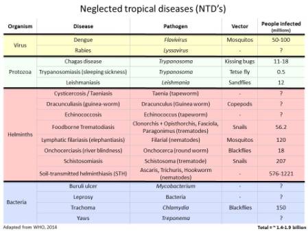
In total the WHO has identified 17 diseases or groups of diseases that fall within this category.


NTD slides from Bonnie Webster.jpg

World Health Organization has identified 17 Neglected Tropical Diseases. 10 of these have been targeted for control and elimination by 2020


The 10 selected by the WHO for control and elimination by 2020 are:


  1. Onchocerciasis (aka river blindness): A blood worm infection transmitted by the bite of infected blackflies causing severe itching and eye lesions as the adult worm produces larvae and leading to visual impairment and permanent blindness.
  2. Dracunculiasis (aka Guinea-worm disease): A roundworm infection transmitted exclusively by drinking-water contaminated with parasite-infected water fleas. The infection leads to meter-long female worms emerging from painful blisters on feet and legs to deposit her young. This leads to fever, nausea and vomiting as well as debilitating secondary bacterial infections in the blisters.
  3. Lymphatic filariasis: A blood & lymph worm infection transmitted by mosquitoes causing abnormal enlargement of limbs and genitals (elephantiasis) from adult worms inhabiting and reproducing in the lymphatic system.
  4. Blinding trachoma: A chlamydial infection transmitted through direct contact with infectious eye or nasal discharge, or through indirect contact (e.g. via flies) with unsafe living conditions and hygiene practices, which if left untreated causes irreversible corneal opacities and blindness. Trachoma is the leading cause of blindness in the word.
  5. Schistosomiasis (aka bilharzia): A blood fluke infection transmitted when larval forms released by freshwater snails penetrate human skin during contact with infested water. The infection leads to anaemia, chronic fatigue and painful urination/defaecation during childhood, later developing into severe organ problems such as liver and spleen damages, bladder cancer, genital lesions and infertility.
  6. Visceral leishmaniasis (aka Kala azar): A protozoan blood parasite transmitted through the bites of infected female sandflies which attacks internal organs which can be fatal within 2 years. 
  7. Soil-transmitted helminths: A group on intestinal worm infections transmitted through soil contaminated by human faeces causing anaemia, vitamin A deficiency, stunted growth, malnutrition, intestinal obstruction and impaired development.
  8. Leprosy: A complex disease caused by infection mainly of the skin, peripheral nerves, mucosa of the upper respiratory tract and eyes.
  9. Chagas disease: A life-threatening illness caused by a blood protozoan parasite, transmitted to humans through contact with vector insects (triatomine bugs), ingestion of contaminated food, infected blood transfusions, congenital transmission, organ transplantation or laboratory accidents.
  10. Human African trypanosomiasis (aka sleeping sickness): A protozoan blood parasitic infection spread by the bites of tsetse flies that is almost 100% fatal without prompt diagnosis and treatment to prevent the parasites invading the central nervous system.


They were selected because the tools to achieve control are already available to us and, for some, elimination should be achievable.


Take the Guinea Worm:


Guinea worm Peter Mayer.jpgGW (SKnopp).jpg

Guinea worm infection - from over 3.5 million people infected in the 80s to less than 130 cases in 2014. Set to be second human disease to be eradicated after smallpox (photo credits David Hamm&Peter Mayer)


In the 1980s over 3.5 million people were infected with Dracunculiasis (i.e. Guinea worm disease), with 21 countries being endemic for the disease. Now, thanks to the global health community efforts and extraordinary support from the Carter Center, only 126 cases were reported in 2014 and only 4 endemic countries remain: Chad, Ethiopia, Mali and South Sudan! If the WHO goal of global eradication of Guinea Worm by 2020 is met then Dracunculiasis is set to become the second human disease in history to be eradicated (the first, and only one, being smallpox). Not bad for an NTD! But there are still challenges!


At the Museum we have a long history of working on health related topics. Indeed our founding father Sir Hans Sloane was a physician who collected and identified plants from all over the world for the purpose of finding health benefits - in fact he developed chocolate milk as a health product.


Today we have a vast and biologically diverse collection of parasites and the insects/crustaceans/snails/arachnids that carry and transmit them. These are used by researchers both in the museum (such as myself and colleagues) but also internationally through collaborative work.


ZEST Zanzibar.jpg

Collaboration is key - Zanzibar Elimination of Schistosomiasis Transmission (ZEST) programme key players: the Zanzibar Ministry of Health, Public Health Laboratories Pemba, the World Health Organization, SCI, SCORE, Swiss TPH, NHM and others


We are immensely proud of our collections and the work we do in this field especially of the biological information we can contribute to health programmes in endemic countries. One of our most exciting contributions is to the Zanzibar Elimination of Schistosomiasis Transmission (ZEST) programme where we are working in collaboration with the Zanzibar Ministry of Health, various NGOs, the World Health Organization and the local communities to identify and implement the best tools and methods to achieve schistosomiasis elimination in Zanzibar. This would be the first time a sub-Saharan African country would achieve schistosomiasis elimination. Fingers-crossed we are up to the challenge! You can read more about this project in an earlier post on our Super-flies and parasites blog


On Thursday we are bringing out our Parasites and Vectors specimens to showcase them to the public galleries and answer any questions relating to these fascinating yet dangerous organisms. Our wonderful scientists and curators will be on hand to talk to people about our collections and research as will collaborating scientists from the London Centre of Neglected Tropical Disease Research who will talk to you about the diseases and the challenges faced to achieve the WHO 2020 goals. Please do pop by and say hello, come and look at our specimens and help us raise awareness of these devastating diseases and the fight to control and eliminate them.


Fieldwork_TZ_FionaAllan_SCAN.jpg

We are working together with schools, communities, government and research institutes to fight Neglected Tropical Diseases. Schistosomiasis fieldwork photo with the team from the National Institute for Medical Research in Tanzania

1

Hello honorary parasitologists,


I know there has been a bit of radio silence on my part and I apologise, summer was calling me and there was a lot of stuff to get through before I could escape on annual leave.


I'm back now and picking up where we left off. Perhaps you are wondering what happened to all those samples we collected in Tanzania in May (see previous posts). Well wonder no longer, I am about to reveal all.


A quick recap of our collecting in Tanzania:

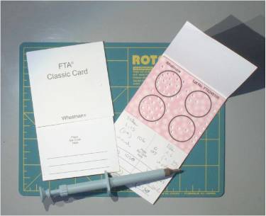
  1. We collected miracidia, the parasite larval stage from infected children, and stored them onto special paper (called Whatman® FTA cards) that stores their genetic material.
  2. We also collected the intermediate host snail from potential transmission sites on the banks of Lake Victoria.
  3. Finally we collected cercariae, the larval stage from infected snails, and stored their genetic material on the Whatman® FTA paper.


Visiting+schools+to+collect+schistosomes700.jpg

Visiting schools to identify infected children, collect the schistosome larval stage (miracidia) and treat the children.


Collecting snails in Tanzania.jpg

Snail collecting on the banks of Lake Victoria.


Schistosoma mansoni cercaraie&Biomphalaria sudanica.jpg

Collecting cercariae, the Schistosoma larval stage, from infected snails.


Whatman FTA and punch.jpg

Whatman® FTA cards store the genetic material of schistosome larvae collected from infected children and/or snails.


Storing our collected schistosome larvae on Whatman® FTA cards is ideal for us because:

  1. We avoid storing them in flammable liquids like ethanol.
  2. We avoid having to bring back live, infected snails.


...two things that aiports and customs really don't like!


The Whatman® FTA cards lyse (break) open the parasite cells and lock the genetic material onto the card, keeping it stable and safe at room temperature until we need to use it. So this means we can bring back our samples safely in our suitcases. Hurrah!


The snails, on the other hand, have to be stored in glass tubes with ethanol, so these we have to leave behind in the safe hands of the National Institute for Medical Research, Mwanza. Our collaborators take good care of them until we can arrange a courier service to bring them to the Museum.


SCAN_FTA card storage closeup.jpg

Whatman® FTA cards with collected schistosome genetic material from Tanzania, catalogued and stored safely by SCAN in the Molecular Collections Facility at the Museum.


Once back in the UK I hand over all collected samples and forms to the wonderful SCAN,Schistosomiasis Collection at the Natural History Museum, team. SCAN takes care of the thousands of schistosome samples collected from all over the world and stored at the Museum. This colleciton is very precious and in high demand for lab-based scienists researching the genome of the parasite and host snail in search of new ways to understand and control the disease. 


SCAN cares for collected samples and manages all the associated data, such as:

  • GPS coordinates - so we know where the sample has come from.
  • Collection method - what technique was used to collect the sample?
  • Date of collection - how old is the sample?
  • Storage medium -  is the sample stored on Whatman FTA card or ethanol or any other storage tool?
  • Data on the parasite host - did the sample come from an infected human, cattle, snail? If a snail what species? If a human what age? Gender? 

And lots more.

Screen shot 2014-08-11 at 20.14.55.jpg

The fieldwork forms I fill in when collecting samples in Tanzania. I hand these forms over to the SCAN team along with all my collected samples. They then have the frustrating task of trying to decipher my handwriting.


You have met two members of the SCAN team in my previous posts; Fiona Allan, who acted as our fieldwork photographer whilst helping me in Tanzania and Muriel Rabone, who came to my rescue after Fiona had to head back to the UK. There is just one more person for you to meet; the ever-patient and resourceful Aidan Emery, who manages SCAN


SCAN_me&F going trhough forms.jpg

Fiona and I going through my fieldwork forms and samples - "what have you written here? It's illegible!" Oops!


As you can see there is A LOT of data that goes with each and every schistosome/snail collected and researchers need to have all this information when analyising a parasite or snail sample. SCAN ensures that all this information is properly entered into a database and linked with the samples stored in the Molecular Collection Facility (more on this in a bit).


The SCAN team has even created a wonderful online catalogue of all the collected samples they care for, along with all the data linked to each sample. This greatly assists researchers from all over the world, allowing them to have a look and see what samples are available to them. 


SCAN_F&A_bluebooties700.jpg

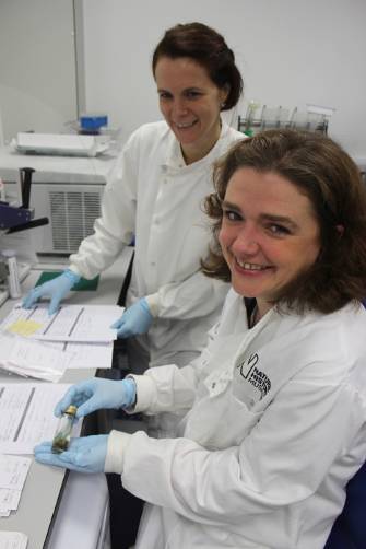
Fiona and Aidan storing the Tanzanian schistosome samples collected onto Whatman® FTA cards. Fiona is showing off the little blue booties we have to wear in the Molecular Collections Facility to avoid bringing in contaminants or anything that could harm the hundreds of thousands of precious samples stored there. 


Screen shot 2014-08-11 at 19.17.56.jpg

The SCAN team takes a photo and measures the size of every snail that arrives from our African collaborators. In order to extract the DNA from the snail for molecular work the shell must be crushed and removed. It is good to have a picture of what the shell looked like before doing so.


The Molecular Collections Facility is a crucial facility in the Museum, as it has all the equipment necessary to keep molecular and genetic samples (such as our schistosome samples) stored safely, stabily and for a long, long time. What equipment am I talking about? I mean: -80 freezers, nitrogen tanks, air-tight cabinets, equipment to release/elute genetic material from stored samples, centrifuges, pipetting robots, you name it. It is run by the very helpful Jackie Mackenzie-Dodds.


MCF_Jackie700.jpg

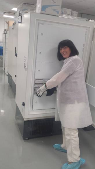
Jackie runs the Molecular Collections Facility where all our schistosome samples are stored. All the freezers and nitrogen tanks have alarms linked to them to make sure they continue to function correctly. If one fails an alarm goes off on Jackie's mobile phone so no matter where she is she is immediately notified and able to respond.


MCF_nitrogen&Jackie.jpg

Jackie is showing me the liquid nitrogen tanks in the Molecular Collections Facility. Whilst nitrogen in gas form is harmless, liquid nitrogen is very, very cold and any contact with it can cause severe frostbite, even freeze your arm off. Also as it boils it uses up a lot of oxygen in the air, which can lead to asphyxiation. So oxygen monitors are always used. Its incredible freezing ability means it is very effective at storing rare, degraded and old tissue samples.


So there you have it, all our samples are archived carefully until we are able to perform the molecular work we need to do for species identification and to determine how the genetic diversity of the parasites is being affected by treatment control programs. My next couple of blood fluke posts will be about the techniques we use to do this. So read up on Polymerase Chain Reactions (PCR)... it does feature!


SCAN_F&me+going+through+forms700.jpg

Fiona and I have managed to decipher my handwriting! Hurrah! The samples are saved!

0

Welcome to the Parasites and Vectors Division blog. Let me introduce our group and the superbugs and parasites we work on (WARNING NASTY IMAGES, strong stomachs required).


The world is full of amazing animals, but there are some that have a more sinister side. Our scientists and curators look at insects, arachnids and worms that live on or inside other animals, including people.


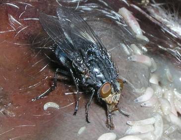
Blue bottle fly - Calliphora vicina - forensic entomology.jpgThe blue bottle fly, Calliphora vicina colonizes corpses and is used in forensic entomology to help crime scene investigators determine time of death.


I’ll be using this blog to write about what we do, why we study these complex organisms and how we collect data in the field and in our laboratories.


I’ll reveal more about the grisly creatures we study later, but for now here’s an introduction to the main players:


  • Flies can cause the horrible disease myiasis, but are also helping scientists to determine crucial information at crime scenes through forensic entomology.
  • Mosquitos have been called the world’s most dangerous animal, carrying diseases like malaria and viruses like dengue.
  • Ticks and mites (Acari) can cause huge damage to crops, and spread diseases such as Lyme disease and babesiosis.
  • Blood flukes are parasitic worms that cause schistosomiasis, a disease affecting over 200 million people worldwide. Museum scientists are studying these worms to help affected countries control schistosomiasis, a neglected tropical disease. More about this in my next post!
  • Flatworms can be parasitic monsters, but their amazing capacity for regenerative growth could inspire regenerative medicine techniques and anti-aging therapies in humans.


Myiasis .jpgMyiasis wounds on sheep in Hungary produced by the spotted flesh ply or screwworm fly (Photo credit Alexander Hall).


We use a range of DNA techniques, from mitogenomics to next generation sequencing to investigate, describe and understand parasitic worms. None of our work would be possible without the Museum’s extensive parasite and vector collections. Erica McAlister curates one of these, the diptera (true flies) collection, which you can read more about on her (very entertaining) blog.


schisto_venous_system_cattle.jpgDon't let size fool you; these tiny blood flukes living in the blood veins of animals cause a debilitating disease called Schistosomiasis.


That’s it for now but check back soon - I’ll be setting off to Tanzania next week in search of blood flukes and will surely have some stories to tell from the field!