
Secondary electron images of volcanic ash from a volcanic plume in the crater rim of Villarrica volcano, Chile, collected using an air filter. Scale bars - 1μm.
The Ultra Plus scanning electron microscope is suitable for high-resolution imaging of biological and non-biological specimens. The microscope's charge compensation system allows non-conducting samples to be imaged without the need for coating.
Key instrument features
- Ultra high-resolution secondary and backscatter electron imaging, utilising in-lens detector technology.
- Energy selective backscatter detector (EsB) features an integrated filtering grid to enhance image quality. It is less sensitive to edge contrast and charging effects, which enables precise imaging and measurement of boundaries, particles and features.
- High-efficiency in-lens secondary electron detector enables high-contrast surface imaging.
- Scanning transmission electron microscope (STEM) detector offers bright field and orientated dark field modes, and a carousel transmission electron microscope (TEM) holder taking up to nine grids.
- Revolutionary charge compensation system for the imaging of non-conducting samples.
- Motorised six-axis super eucentric stage (-4° to 70° tilt), coupled with Zeiss SmartSEM image navigation, enables quick and simple sample navigation.
- Energy dispersive X-ray (EDX) micro-analysis.
Example applications
-
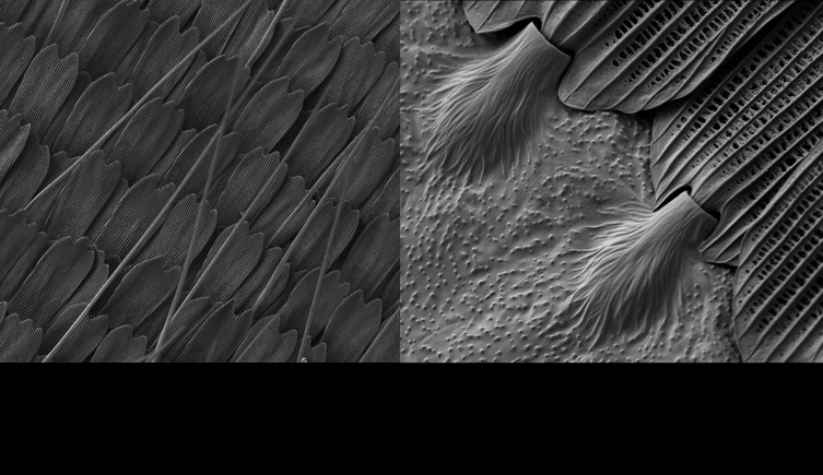
Secondary electron images of butterfly scales from Artogeia napi. Acquired using the Zeiss Ultra Plus charge compensation system and low kV.
-

Secondary electron images of butterfly scales from Vanessa atalanta. Acquired using the Zeiss Ultra Plus charge compensation system and low kv.
-

Carbon nanotubes imaged using the in-lens secondary detector at low kv.
-

Copper oxide nanoparticles imaged using the scanning transmission electron microscope detector in dark field mode.
-

High-resolution energy dispersive X-ray phase map of the Murchison meteorite prepared using Oxford Instruments X-Max silicon drift detector and INCA automated montaging software. Red - magnesium, blue - aluminium, green - calcium.
About EDX analysis
Energy dispersive X-ray spectroscopy (EDX) is a kind of scanning electron microscopy that involves X-ray micro-analysis. The key features of this technique are:
- Non-destructive qualitative analysis can be carried out for elements with an atomic number greater than five.
- Detection limits are of the order of 0.2 weight per cent, dependent on the type of specimen, elements of interest, etc.
- Analyses can be undertaken in spot mode or the beam can be scanned to acquire X-ray element maps of areas of the sample surface.
- Automated image and elemental analysis can be carried out over large specimen areas using Oxford Instruments INCA software's feature mode.
- Backscattered electron and X-ray element map montages can be acquired and automatically stitched using Oxford Instruments INCA software's montage mode.
Key facts
Techniques: high-resolution SEM imaging with X-ray micro-analysis
Magnifications: 12-1,000,000x for secondary electron (SE) images, 100-1,000,000x for backscattered electron (BSE) images
Resolution: 1nm, dependent on working conditions
Image output: 8-bit and 16-bit TIFFs, up to 3072 x 2304 pixels
Detectors
- four-quadrant solid state backscattered detector (AsB)
- conventional secondary electron detector (Everhardt-Thornley)
- in-lens secondary electron detector
- in-lens energy and angle selective backscatter detector (EsB)
- multimode scanning transmission electron microscope (STEM) detector
- energy dispersive X-ray (EDX) detector (Oxford Instruments X-Max detector)