
Hello Super-flies and Parasites fans!
We are back with all things nasty from the Parasites and Vectors division here at the Museum. There have been some exciting developments in the New Year, most importantly the launch of the Museum’s brand new website!
This is another ‘Forever Flies’ series of blog posts, bringing you news from the Museum'sforensic entomologygroup.

Forever Flies is our forensic entomology blog series. This image shows a carrion-eating greenbottle blowfly.
Forensic Entomology
You will remember from my previous Forever Flies post that forensic entomology is the study of the insects and arthropods found at a crime scene. The most common role for Museum forensic entomologists is establishing a minimum time since death in suspicious cases, by analysing the carrion insects on the body.


Blowflies use the bodies of dead animals to grow and develop. The rate at which they do this, going from egg to larva to pupa to adult fly, is pretty consistent and depends largely on ambient temperature. Forensic entomologists use this to determine the minimum post-mortem interval (PMImin), which helps crime scene investigators determine approximate time-of-death.
Thanks to entomological expertise (Greek – entomo = insect, logos = knowledge) scientists can collect insects from a corpse and/or crime scene, determine what stage in their life cycle the insects have reached and, using their knowledge on the duration of each stage of the insects’ life cycle, determine how long ago the parent insect laid her eggs on the corpse.
This gives an incredibly useful estimate of the minimum amount of time this body has been dead (minimum post-mortem interval - PMImin), which helps crime scene investigators determine approximate time-of-death. The more accurate this minimum post-mortem interval is, the more accurate the time of death can be. Knowing time of death can focus the police investigation and suggest the likelihood of a suspect’s involvement.
Scientists can also use these insects to determine if the body has been moved since death and how long a body was exposed above ground before burial.
Metamorphosis in pupae
Flies spend over 50% of their developmental life in the pupae stage, protectively encased inside a hard shell (called a puparium) where they slowly transform from a maggot into a fly in a process called metamorphosis (Greek again - Meta = change, morphe = form).
A puparium looks quite bland and boring but underneath there are all sorts of wonderful things going on. Scientists can remove the shell and, using traditional microscopy, take a look at the fascinating changes of metamorphosis. But this process does destroy the pupa sample, making it difficult to work out how long it takes for the pupa to go through the different stages of metamorphosis.
Scientists know that the length of time metamorphosis takes to complete really depends on temperature, the question is can we use our knowledge of the process to pinpoint a more accurate estimate of PMImin? What forensic scientists need is a standardised method to work out:
- At what stage in the metamorphosis process is the pupa
- how long did it take to reach this stage
If these two points can be determined then scientists can provide a far more accurate PMImin.
The ‘MORPHIC’ project
Dr Daniel Martin-Vega, a forensic entomologist, has joined the Museum from the University of Alcalá in Spain to research carrion fly pupae and to develop a standardised protocol for aging pupae (as in determining their age) that can be used by forensic scientists. This project is called MORPHIC and is funded by the European Commission through a Marie-Curie fellowship.
It sounds all neat, logical and tidy but there is A LOT of work and dedication involved!
For this projectDaniel is raising two species of the carrion-loving blowflies, the greenbottle blowfly Lucilia sericata and the bluebottle blowfly Calliphora vicina. The flies live in netting covered cages, where they feed and reproduce whilst he monitors them.
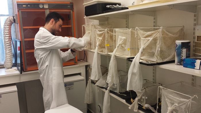 Daniel showing me the Diptera (insect) culture room. Each netting-covered box has a species of carrion blowfly in it. He is researching the pupae of these flies to see if he can improve the estimate of PMImin and thus improve the information given to crime scene investigators.
He also has to collect the post-feeding maggots and place them in a box with some nice clean soil for them to happily grow until they are ready to start the metamorphosis process. These boxes are then placed in a cabinet kept at a specific temperature. Since the rate of metamorphosis largely depends on temperature it is very important the Daniel can control this environmental factor in order to document the rate of change at different temperatures.

The maggot house! This is a comfy box with soil where maggots crawl around and prepare to pupate. When the maggots start pupating Daniel has to come in every 6 hours or so to monitor and collect them for his research

Blowfly maggots and pupae.
Once the maggots start to pupate Daniel has to collect the pupae:
I come in every 6 hours when the maggots start to pupariate in order to collect blowfly pupae at 6-hour intervals during the first 48 hours after puparium formation (the period when the greatest morphological changes of metamorphosis occur). Luckily, I only do this from time to time. After that, the collection of pupae is just daily until the adult flies’ emergence.

Daniel sieving out the pupae from the box.

Maggots and pupae, oh my!
Watch those maggots wriggle about!
He then has to sieve out the pupae from the soil and carefully place them in a petridish labelled with the blowfly species name, the date collected and the time collected. These petridishes are also placed in the special temperature-control cabinet.

Daniel has separated out the pupae of different species of blowfly. Each petridish with pupae has the species name, the date collected and the time collected.
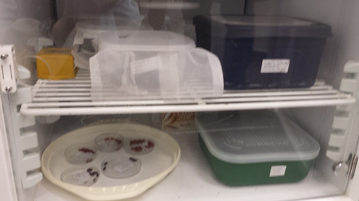
The petridishes are kept at a specific temperature. Since the rate of metamorphosis largely depends on temperature it is very important the Daniel can control this environmental factor.
Daniel uses the Museum’s wonderful micro-computed tomography (micro-CT) scanner to take detailed images of the inside of the pupae without destroying them. A micro-CT scanner is a type of X-ray scanner that produces 3D images, much like a hospital CAT scanner, but at a much smaller scale and a higher resolution. The results are like 3D microscope images!


Daniel with colleague Dr Thomas Simonsen using the Museum’s micro-CT scanner to look at 3D images of blow-fly pupae. The micro-CT scanner uses x-ray technology to produce 3D 'microscopy' images at high resolution without damaging the sample.
By using the Museum’s micro-CT scanner Daniel can take these detailed images at specific time points of the metamorphosis process. He will then have a catalogue of images of the blow fly pupal development at specific temperatures. This catalogue of images will be used to develop a standardised tool to determine the age of blow fly pupae. Then when pupae are collected from a crime scene, they can be compared to this catalogue and scientists will be able to determine how long the fly has been in its pupal stage. Giving scientists a more accurate estimate of PMImin! Ta daaaaa!


Micro-CT scanner images of a bluebottle blowfly Calliphora vicina pupa. The one on the left is at 48 hours, the one on the right at 216 hours. You can see the difference in development between the two pupa images.

Dorsal micro-CT scanner image of a blowfly pupa.
I hope you enjoyed this post. If you fancy a stab at a bit of CSI work why not check out the Museum's Crime Scene Live After Hours events.